. 2002 Aug 6;99(16):10299-304.
doi: 10.1073/pnas.162352799. Epub 2002 Jul 22.

Severe block in processing of proinsulin to insulin accompanied by elevation of des-64,65 proinsulin intermediates in islets of mice lacking prohormone convertase 1/3

Affiliations

Severe block in processing of proinsulin to insulin accompanied by elevation of des-64,65 proinsulin intermediates in islets of mice lacking prohormone convertase 1/3

Xiaorong Zhu et al. Proc Natl Acad Sci U S A. .

Abstract

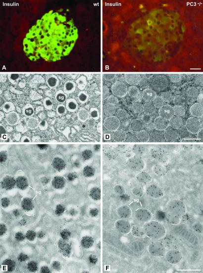
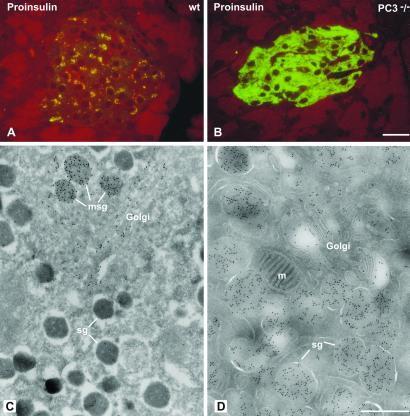
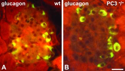
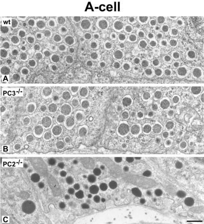
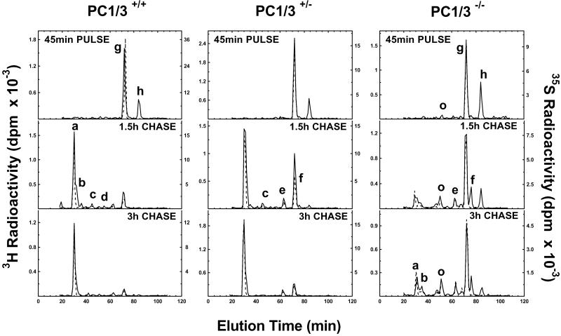
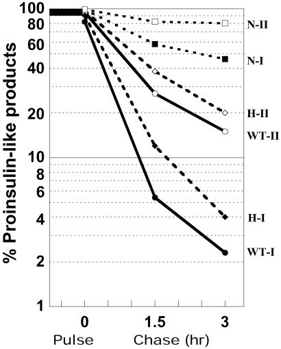
The neuroendocrine processing endoproteases PC2 and PC1/3 are expressed in the beta cells of the islets of Langerhans and participate in the processing of proinsulin to insulin and C-peptide. We have previously shown that disruption of PC2 (SPC2) expression significantly impairs proinsulin processing. Here we report that disruption of the expression of PC1/3 (SPC3) produces a much more severe block in proinsulin conversion. In nulls, pancreatic and circulating proinsulin-like components comprise 87% and 91%, respectively, of total insulin-related immunoreactivity. Heterozygotes also show a more than 2-fold elevation in proinsulin levels to approximately 12%. Immunocytochemical and ultrastructural studies of the beta cells reveal the nearly complete absence of mature insulin immunoreactivity and its replacement by that of proinsulin in abundant immature-appearing secretory granules. In contrast, alpha cell morphology and glucagon processing are normal, and there is also no defect in somatostatin-14 generation. Pulse-chase labeling studies confirm the existence of a major block in proinsulin processing in PC1/3 nulls with prolongation of half-times of conversion by 7- and 10-fold for proinsulins I and II, respectively. Lack of PC1/3 also results in increased levels of des-64,65 proinsulin intermediates generated by PC2, in contrast to PC2 nulls, in which des- 31,32 proinsulin intermediates predominate. These results confirm that PC1/3 plays a major role in processing proinsulin, but that its coordinated action with PC2 is necessary for the most efficient and complete processing of this prohormone.


Figures

Fig 1.
Fig 1.
Insulin immunoreactivity is decreased in PC3−/− mice. (A and B) Paraffin sections of pancreatic islets of wild-type (wt) (A) and knockout (B) mice incubated with the anti-insulin antibody by the immunofluorescence method. In control islet (A) the mass of insulin B cells is brightly immunostained, whereas in PC3−/− islet (B), the insulin cells show a very strong reduction in labeling. The reduced level of insulin immunoreactivity was also detectable on thin sections labeled with insulin antibody revealed by anti-mouse IgG-gold. The quantitation indicated 502 ± 24 gold particles/μm2 of 20 secretory granules (sg, E) versus 169 ± 9 gold particles/μm2 (PC3−/− mice, F). The secretory granules in PC3−/− mice also had a different morphology characterized by a pale content and a thin halo (D), as compared with the characteristic dense core and wide halo in the control wt mice (C). (Bars: A and B, 20 μm; CF, 0.5 μm.)
Fig 2.
Fig 2.
Proinsulin immunoreactivity is increased in PC3−/− mice. (A and B) Staining by immunofluorescence for proinsulin on semithin Epon sections from wild-type (wt) and mutant mice. In wt islets proinsulin staining has the characteristic Golgi-like perinuclear distribution (A); in mutant PC3−/− mice, proinsulin staining is abundant throughout the entire cytoplasm (B). Proinsulin labeling at the ultrastructural level is restricted to the Golgi complex and the maturing secretory granules (msg) in wt mice (C). In PC3−/− mice, gold labeling is present over the Golgi complex and the entire population of secretory granules (sg) (D). m = Mitochondrion. (Bars: A and B, 20 μm; C and D, 0.5 μm.)
Fig 3.
Fig 3.
Glucagon immunoreactivity shows a similar pattern of distribution in wild-type (wt) and PC3 null mice. Shown are paraffin sections of pancreatic islets from wild-type (A) and PC3 null (B) mice incubated with C-terminal anti-glucagon antibody by the immunofluorescence method. The amount and distribution of immunoreactive cells are similar in wt and null islets. (Bar: 20 μm.)
Fig 4.
Fig 4.
The morphology of glucagon cell secretory granules is unaltered in PC3 null mice. Shown are thin section electron micrographs comparing the morphologic appearance of α cell secretory granules in wild-type (wt) (A), PC3(−/−) (B), and PC2(−/−) (C) mice. PC3 null and wt secretory granules show a similar aspect, characterized by a dense granule core separated by a distinct clear halo from the limiting membrane. By contrast, in PC2 null glucagon cell granules, the halo is absent and the denser content reaches the granule limiting membrane. (Bar: 0.5 μm.)
Fig 5.
Fig 5.
Conversion of proinsulin to insulin in pulse–chase metabolic labeling. Islets from wild-type (Left), heterozygous (+/−) (Center), or null (−/−) (Right) mice were labeled with 35S-Met (dashed lines) or 3H-Leu (solid lines) in high glucose for 45 min and then chased in low glucose for a total of 3 h. Proteins were extracted from islets at indicated times, immunopurified, and resolved on HPLC, as described in Materials and Methods. Peaks are identified (see ref. 12) as follows: a = mouse insulin II; b = mouse insulin I; c and d = des-31,32 mProinsulins II and I, respectively; e and f = des-64,65 mProinsulins II and I, respectively; g and h = intact mProinsulins II and I, respectively; and o = oxidized mProinsulin II. Note the rapid conversion of both proinsulins I and II in wild-type islets, whereas conversion is clearly slowed in heterozygous islets at 1.5 h and markedly reduced throughout the chase period in null islets, accompanied by the appearance of des-64,65 intermediate peaks (e and f).
Fig 6.
Fig 6.
Time course of accumulation of des-64,65 proinsulin intermediates in wild-type (+/−) and null islets during a 3-h chase after a 45-min pulse. Note much greater accumulation in nulls of both proinsulin I and II intermediates. The much greater production of the des-64,65 intermediate I is likely caused by the presence of a P4 arginine residue at position 62 in proinsulin I rather than glutamine, as in proinsulin II. ◊, Wild type; ○, heterozygotes; •, nulls. Dashed line denotes des- 64,65 proinsulin I and solid line indicates des-64,65 proinsulin II.
Fig 7.
Fig 7.
Semilog plot of the disappearance rates of proinsulin-like components during a 3-h chase in islets of PC1/3 (+/+), (+/−), and (−/−) mice. Black symbols denote proinsulin I, and white symbols denote proinsulin II components. N, null; H, heterozygous; WT, wild type.

References

    1. Steiner D., Chan, S. & Rubenstein, A. (2000) in Handbook of Physiology, The Endocrine System, eds. Jefferson, L. & Cherrington, A. (Oxford Univ. Press, New York), Vol. II, pp. 49–77.
    1. Steiner D. (2001) in The Enzymes, eds. Dalbey, R. E. & Sigman, D. S. (Academic, New York), Vol. XXII, pp. 163–198.
    1. Goodge K. A. & Hutton, J. C. (2000) Semin. Cell. Dev. Biol. 11, 235-242. - PubMed
    1. Zhou A., Webb, G., Zhu, X. & Steiner, D. F. (1999) J. Biol. Chem. 274, 20745-20748. - PubMed
    1. Seidah N. G. & Chrétien, M. (1999) Brain Res. 848, 45-62. - PubMed

Publication types