

In vivo quantification of localized neuronal activation and inhibition in the rat brain using a dedicated high temporal-resolution beta +-sensitive microprobe
- PMID: 12136134
- PMCID: PMC125052
- DOI: 10.1073/pnas.162368899
In vivo quantification of localized neuronal activation and inhibition in the rat brain using a dedicated high temporal-resolution beta +-sensitive microprobe
Abstract
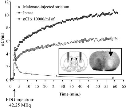
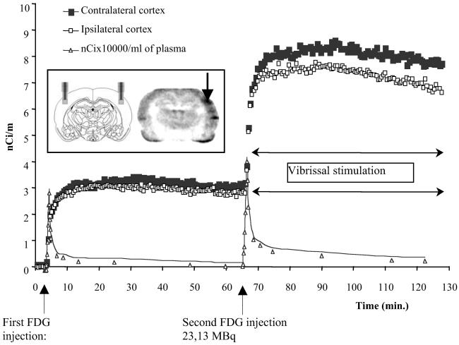
Understanding brain disorders, the neural processes implicated in cognitive functions and their alterations in neurodegenerative pathologies, or testing new therapies for these diseases would benefit greatly from combined use of an increasing number of rodent models and neuroimaging methods specifically adapted to the rodent brain. Besides magnetic resonance (MR) imaging and functional MR, positron-emission tomography (PET) remains a unique methodology to study in vivo brain processes. However, current high spatial-resolution tomographs suffer from several technical limitations such as high cost, low sensitivity, and the need of restraining the animal during image acquisition. We have developed a beta(+)-sensitive high temporal-resolution system that overcomes these problems and allows the in vivo quantification of cerebral biochemical processes in rodents. This beta-MICROPROBE is an in situ technique involving the insertion of a fine probe into brain tissue in a way very similar to that used for microdialysis and cell electrode recordings. In this respect, it provides information on molecular interactions and pathways, which is complementary to that produced by these technologies as well as other modalities such as MR or fluorescence imaging. This study describes two experiments that provide a proof of concept to substantiate the potential of this technique and demonstrate the feasibility of quantifying brain activation or metabolic depression in individual living rats with 2-[(18)F]fluoro-2-deoxy-d-glucose and standard compartmental modeling techniques. Furthermore, it was possible to identify correctly the origin of variations in glucose consumption at the hexokinase level, which demonstrate the strength of the method and its adequacy for in vivo quantitative metabolic studies in small animals.
Figures
References
-
- Sokoloff L., Reivich, M., Kennedy, C., DesRosiers, M. H., Patlak, C. S., Pettigrew, K. D., Sakurada, D. & Shinohara, M. (1977) J. Neurochem. 28, 897-916. - PubMed
-
- Weber D. A., Ivanovic, M., Franceschi, D., Strand, S. E., Erlansson, K., Franceschi, M., Atkins, H. L., Coderre, J. A., Susskind, H., Button, T. & Ljunggren, K. (1994) J. Nucl. Med. 35, 342-348. - PubMed
-
- Lecomte R., Cadorette, J., Rodrigue, S., Lapointe, D., Rouleau, D., Bentourkia, M., Yao, R. & Msaki, P. (1996) IEEE Trans. Nucl. Sci. 43, 1952-1957.
-
- Watanabe M., Okada, H., Shimizu, K., Omura, T., Yoshikawa, E., Kosugi, T., Mori, S. & Yamasmita, T. (1996) IEEE Trans. Nucl. Sci. 2, 1330-1334.
-
- Ziegler S. I., Pichler, B., Boening, G., Rafecas, M., Pimpl, W., Lorenz, E., Schimtz, N. & Schwaiger, M. (2001) Eur. J. Nucl. Med. 28, 136-143. - PubMed
Publication types
MeSH terms
Substances
LinkOut - more resources
Full Text Sources
