

Upregulated eotaxin expression and T cell infiltration in the basal and papillary epithelium in cows' milk associated reflux oesophagitis
- PMID: 12138061
- PMCID: PMC1719188
- DOI: 10.1136/adc.87.2.124
Upregulated eotaxin expression and T cell infiltration in the basal and papillary epithelium in cows' milk associated reflux oesophagitis
Abstract
Background: Cows' milk sensitive reflux oesophagitis is an emerging clinical entity in children, normally indistinguishable from primary gastro-oesophageal reflux (GOR) apart from the response to dietary antigen exclusion. It is conjectural whether a tendency towards mucosal eosinophilia distinguishes this group from primary GOR.
Aims: To determine whether there may be differences in the mucosal lesion within the oesophagus in those children with reflux in association with cows' milk induced small bowel pathology, particularly in relation to the eosinophil chemokine eotaxin.
Methods: A total of 29 children underwent endoscopic assessment, including nine with cows' milk sensitive enteropathy (CMSE) and associated GOR, seven histologically normal controls, six with primary GOR, and seven disease controls. Oesophageal biopsy specimens were examined immunohistochemically for the chemokines eotaxin and MCP-2, and T cell lineage and activation markers.
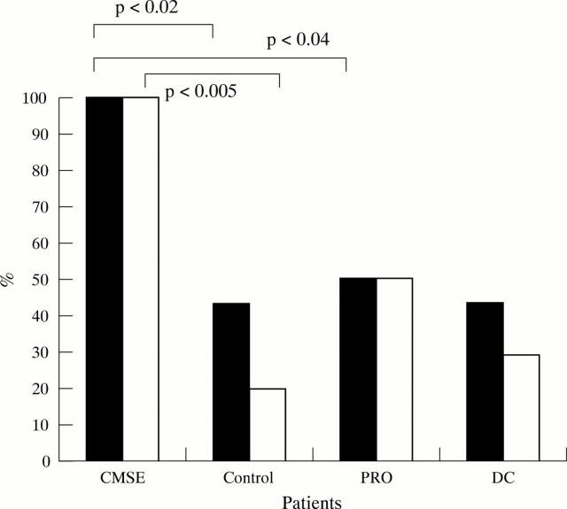
Results: Strong upregulation of eotaxin expression, limited to basal and papillary epithelium, occurred in all CMSE patients. By contrast, weak expression was seen in a minority of controls and in 50% of primary GOR patients. Infiltration of CD3, CD4, and CD8 lymphocytes occurred in similar distribution in CMSE patients, significantly increased above controls. Significant upregulation of activation markers (CD25, HLA-DR) was also seen in the CMSE group within basal and papillary epithelium compared to controls and primary GOR.
Conclusion: Basal and papillary epithelial eotaxin expression, with focal lymphocyte activation, was seen in infants with CMSE associated GOR. This preliminary study provides early evidence to suggest a pathogenesis distinct from primary GOR, in which specific recruitment of T cells and eosinophils may contribute to oesophageal dysmotility.
Figures


References
Publication types
MeSH terms
Substances
LinkOut - more resources
Full Text Sources
Research Materials
Miscellaneous