. 2002 Aug;22(16):5698-707.
doi: 10.1128/MCB.22.16.5698-5707.2002.

Regulation of alternative splicing by the ATP-dependent DEAD-box RNA helicase p72

Affiliations

Regulation of alternative splicing by the ATP-dependent DEAD-box RNA helicase p72

Arnd Hönig et al. Mol Cell Biol. 2002 Aug.

Abstract

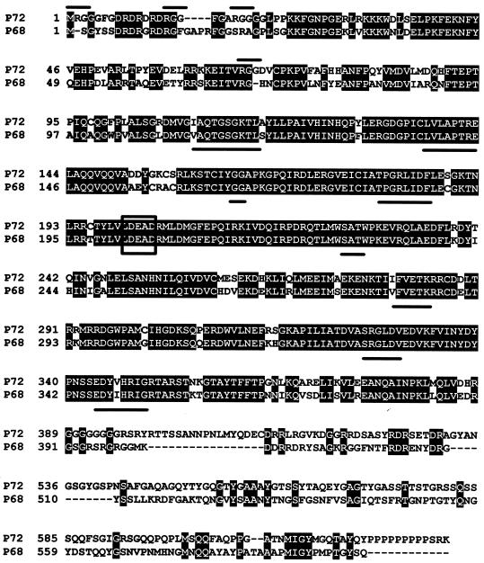
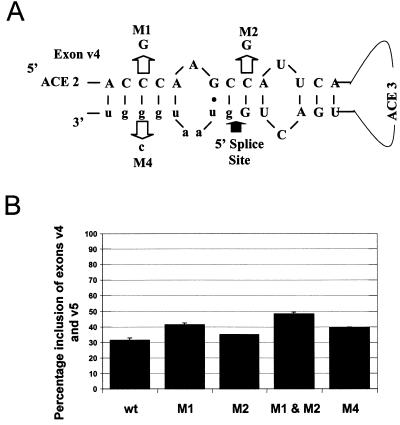
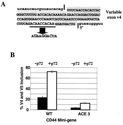
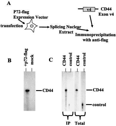
Although a number of ATP-dependent RNA helicases are important for constitutive RNA splicing, no helicases have been implicated in alternative RNA splicing. Here, we show that the abundant DEAD-box RNA helicase p72, but not its close relative p68, affects the splicing of alternative exons containing AC-rich exon enhancer elements. The effect of p72 was tested by using mini-genes that undergo different types of alternative splicing. When the concentration of p72 was increased in transient transfections, the inclusion of enhancer-containing CD44 alternative exons v4 and v5 increased using a mini-gene that contained these exons and their flanking introns inserted into a beta-globin gene. Other types of alternative splicing were not impacted by altering p72 concentrations. Mutation of the p72 helicase ATP-binding site or deletion of the carboxy-terminal region of the protein reduced the ability of the transfected protein to affect CD44 variable exon splicing. Use of in vitro extracts overexpressing p72 indicated that p72 becomes associated with complexes containing precursor RNA. Helicases have been implicated both in altering RNA-RNA interactions and in remodeling RNA-protein complexes. CD44 exon v4 contains a potential internal secondary structure element that base pairs the 5' splice site with a region inside the exon located between enhancer elements. Mutations that destroyed this complementarity modestly increased inclusion in the absence of p72 but still responded to increasing p72 concentration like the wild-type exon, suggesting that p72 might have effects on protein-RNA interactions. In agreement with this hypothesis, p72 was not able to restore the inclusion of an exon mutated for its major enhancer element. Our results suggest that RNA helicases may be important alternative splicing regulatory factors.


Figures

FIG. 1.
FIG. 1.
Comparison of the human p68 and p72 ATP-dependent DEAD-box RNA helicases. Identical amino acids are shaded in black. The DEAD box is boxed, conserved sequences common to the ATP-dependent helicases are underlined, and RGG sequences unique to p72 are overlined. Alignment was provided by ALIGN and BOXSHADE.
FIG. 2.
FIG. 2.
Increasing the in vivo concentration of p72 RNA helicase increases inclusion of CD44 variable exons v4 and v5. (A) CD44 alternative splicing. In diagram 1, the exon structure of the human CD44 gene is depicted. Constitutive exons are shaded black, and alternative exons (termed variable exons) are shaded gray. In diagram 2, the employed CD44 mini-gene in which CD44 variable exons v4 and v5 and their surrounding intron sequences have been inserted into a β-globin mini-gene driven from the CMV promoter is depicted. Globin exons are shaded white, and CD44 exons are shaded gray. In diagram 3, the sequence of CD44 exon v4 is shown. Exon sequences are capitalized; intron sequences are lowercase. The AC-rich exon enhancer (ACE) is underlined. (B) Transfection of p72 or p68. The mini-gene shown in panel A was cotransfected into HeLa cells with 0, 1, 2, or 4 μg of an expression plasmid coding for full-length human p72 (lanes 1 through 4) or human p68 (lanes 5 through 8). RNA splicing patterns were characterized by RT-PCR amplification of total cell RNA with primers specific for the exons flanking the CD44 variable exons. Bands corresponding to the inclusion of no CD44 exons (the skip product), one CD44 variable exon (exons v4 and v5 are of almost identical length), or both CD44 variable exons are indicated by the shading in of the boxes between the gels. The identity of each band was confirmed by sequencing of PCR products. Numbers below the gels and lane numbers indicate the percentage of product RNA containing both exons v4 and v5 as determined by scanning of gels in a phosphorimager. wt, wild type.
FIG. 3.
FIG. 3.
Maximum stimulation of CD44 alternative splicing required multiple domains within p72. Deletion and point mutants of p72 were prepared and assayed by cotransfection with the CD44 mini-gene. (A) Diagram of the constructed mutations. Domains of p72 are indicated by shaded boxes. Point mutations are indicated by the residue number of the mutation and the alteration. p300 was a translational stop mutation introduced at amino acid 300, and p437 was a truncation mutant at amino acid 437 lacking the SG-rich C-terminal tail of the protein. All mutants contained an N-terminal Flag tag equivalent to that present in the wild-type protein. (B and D) Quantification of the ability of each mutant protein to affect the inclusion of CD44 variable exons v4 and v5. The percentage of RNA resulting from the inclusion of variable exons v4 and v5 was calculated from phosphorimager tracing of a gel of RT-PCR amplification of RNAs from cotransfections of the CD44 mini-gene and various forms of p72 as described in the legend to Fig. 2. Standard deviations from multiple experiments are indicated. The last bar indicates the level of the inclusion in the absence of cotransfected p72. wt, wild type. (C) Western blot of total cell protein following transfection with the indicated p72 genes. Detection was with the anti-Flag antibody.
FIG. 4.
FIG. 4.
A stem loop created by base pairing of exon sequences to a region of the intron immediately downstream of the exon v4 5′ splice site may function to minimize exon inclusion. (A) Diagram of a potential stem loop at the 3′ end of exon 4. Base pairing and introduced mutations that should disrupt the stem are indicated. Exon sequences are in capitals, and intron sequences are in lowercase. Two of the AC-rich elements (ACE 2 and ACE 3) are indicated, as is the 5′ splice site. (B) Levels of inclusion of exons v4 and v5 of the mutants in the absence of cotransfecting p72. Transfections and quantification are as described in the legend to Fig. 3.
FIG. 5.
FIG. 5.
The effect of p72 is dependent upon a wild-type exon enhancer within CD44 variable exon v4. (A) The sequence of CD44 exon v4 including the AC-rich enhancer (ACE) and a mutant version of the enhancer (ACE 3). The wild-type and ACE 3 mutant mini-genes were cotransfected with wild-type p72, and RNA splicing phenotypes were assayed by RT-PCR as described in the legend to Fig. 2. The mutant produces RNA including exon v5 but not exon v4 (41). (B) Quantification of the ability of p72 to produce RNA that includes exons v4 and v5. Quantification was performed as described in the legend to Fig. 3.
FIG. 6.
FIG. 6.
Increasing the concentration of p72 had minimal impact on the alternative splicing of other pre-mRNAs capable of splicing choices. Wild-type p72 was cotransfected with mini-genes containing alternative exons 4 through 6 from the human CT/CGRP genes, a constructed adenovirus gene with a first exon containing two 5′ splice sites, and a construct containing a weakened β-globin internal exon. (A) The utilized CT/CGRP gene has exons 4 through 6 of the human CT/CGRP gene and their flanking introns fused to exon 1 from the adenovirus major late transcription unit. Alternative splicing of the mini-gene results in two polyadenylated (An) products: one ending in exon 4 and one resulting from the skipping of exon 4 and inclusion of exons 5 and 6. Alternative products were detected by simultaneous RT-PCR with oligonucleotides (a, b, and c) specific for exons 4 and 5 (26). The effects of p72 were compared to those of cotransfection with the protein YB-1, which has an effect on CD44 alternative splicing (41). Product bands resulting from the usage or skipping of exon 4 are indicated. The percentage of product RNA resulting from the inclusion of exon 4 as determined in the phosphorimager is shown below the gel. (B) The diagrammed mini-gene consisting of sequences from the second exon of the major late adenovirus transcription unit was cotransfected with p72. RNA products resulting from the use of the proximal and distal 5′ splice site are indicated. (C) The diagrammed mini-gene was a derived β-globin gene containing an internally deleted internal exon previously shown to be impaired for inclusion in HeLa cells. RNA products resulting from inclusion or skipping of the middle exon are indicated.
FIG. 7.
FIG. 7.
p72 associates with precursor RNA in in vitro splicing extracts. (A) HeLa cells were transfected with an expression vector for p72-Flag. Standard splicing extract was prepared from the nuclei of these cells. Radiolabeled substrate RNAs containing a portion of the CD44 exon v4, which was comprised of AC-rich sequences or a control, were added to the extract and incubated under splicing conditions for 10 min. Radiolabeled RNA was immunoprecipitated with the anti-Flag antibody. (B) Immunoprecipitation of CD44 RNA sequences from extract made from cells expressing tagged p72 versus mock-transfected cells is shown. Equal amounts of an RNA containing CD44 exon v4 sequences were added to extracts made from p72- or mock-transfected HeLa cells. After a 10-min incubation under splicing conditions, reactions were immunoprecipitated with anti-Flag antibodies and immunoprecipitated RNAs were displayed on a denaturing gel. (C) Comparison of the ability of p72 to associate with CD44 exon 4 or control RNA sequences is shown. Equal amounts of CD44 or a control RNA (Total lanes) were incubated with the extract expressing p72-Flag and immunoprecipitated (IP lanes) with anti-Flag antibodies after 10 min of incubation. Immunoprecipitated RNAs are indicated.

Similar articles

Cited by

References

    1. Arning, S., P. Gruter, G. Bilbe, and A. Kramer. 1996. Mammalian splicing factor SF1 is encoded by variant cDNAs and binds to RNA. RNA 2:794-810. - PMC - PubMed
    1. Caruthers, J. M., and D. B. McKay. 2002. Helicase structure and mechanism. Curr. Opin. Struct. Biol. 12:123-133. - PubMed
    1. Chen, J. Y., L. Stands, J. P. Staley, R. R. Jackups, L. J. Latus, and T. H. Chang. 2001. Specific alterations of U1-C protein or U1 small nuclear RNA can eliminate the requirement of Prp28p, an essential DEAD box splicing factor. Mol. Cell 7:227-232. - PubMed
    1. de la Cruz, J., D. Kressler, and P. Linder. 1999. Unwinding RNA in Saccharomyces cerevisiae: DEAD-box proteins and related families. Trends Biochem. Sci. 24:192-199. - PubMed
    1. Dominski, Z., and R. Kole. 1992. Cooperation of pre-RNA sequence elements in splice site selection. Mol. Cell. Biol. 12:2108-2114. - PMC - PubMed

Publication types

MeSH terms

LinkOut - more resources