. 2002 Aug;22(16):5708-20.
doi: 10.1128/MCB.22.16.5708-5720.2002.

Steady-state nuclear localization of exportin-t involves RanGTP binding and two distinct nuclear pore complex interaction domains

Affiliations

Steady-state nuclear localization of exportin-t involves RanGTP binding and two distinct nuclear pore complex interaction domains

Scott Kuersten et al. Mol Cell Biol. 2002 Aug.

Abstract

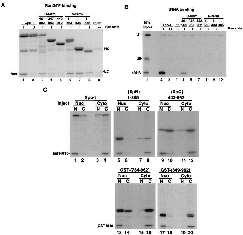
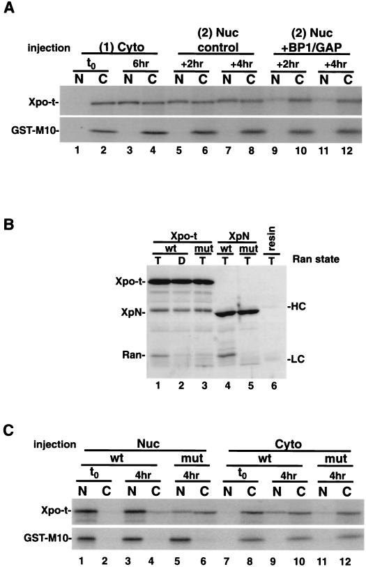
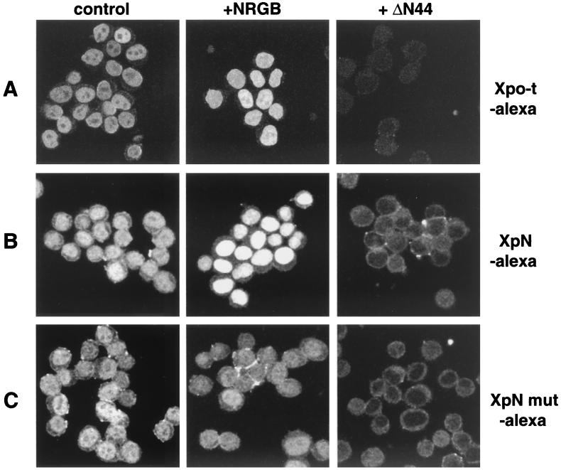
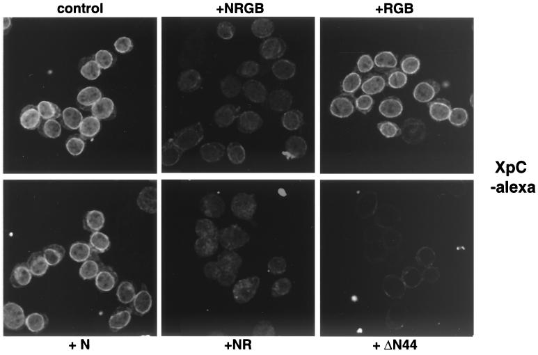
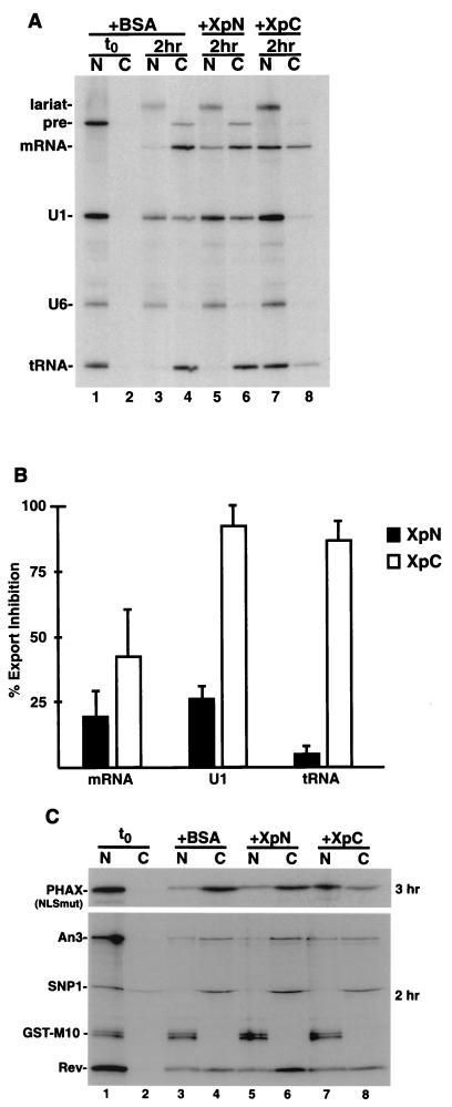
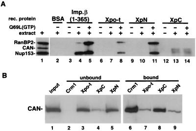
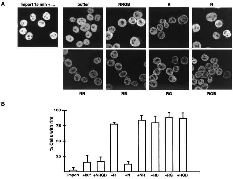
Vertebrate tRNA export receptor exportin-t (Xpo-t) binds to RanGTP and mature tRNAs cooperatively to form a nuclear export complex. Xpo-t shuttles bidirectionally through nuclear pore complexes (NPCs) but is mainly nuclear at steady state. The steady-state distribution of Xpo-t is shown to depend on its interaction with RanGTP. Two distinct Xpo-t NPC interaction domains that bind differentially to peripherally localized nucleoporins in vitro are identified. The N terminus binds to both Nup153 and RanBP2/Nup358 in a RanGTP-dependent manner, while the C terminus binds to CAN/Nup214 independently of Ran. We propose that these interactions increase the concentration of tRNA export complexes and of empty Xpo-t in the vicinity of NPCs and thus increase the efficiency of the Xpo-t transport cycle.


Figures

FIG. 1.
FIG. 1.
In vitro binding and shuttling activities of full-length Xpo-t and Xpo-t with N- and C-terminal truncations. (A) RanGTP binding. zzXpo-t and the truncation mutants indicated were expressed in E. coli and prebound to IgG-Sepharose, mixed with recombinant RanGTP (T, lanes 1 and 3 to 8) or RanGDP (D, lane 2), and incubated at room temperature while being rotated for 1 h. The resin was collected and washed three times with ice-cold binding buffer, and bound proteins were eluted with sample buffer. Samples were separated by SDS-12% PAGE and stained with Coomassie blue. RanGTP can bind to Xpo-t (lane 1) and all three N-terminal fragments of Xpo-t (lanes 6 to 8). In addition, removal of the first 45 amino acids of Xpo-t (lane 3) does not eliminate Ran-binding activity; however, mutants with further deletions (lanes 4 and 5) no longer bind. HC and LC, background elution of the heavy and light chains from the IgG-Sepharose. (B) tRNA binding. A mixture of U1ΔSm, U6Δss, and yeast tRNAPhe (lane 1) was incubated with zzXpo-t or the indicated truncation mutants in the presence of RanGTP (lanes 2 and 5 to 10), RanGDP (lane 3), or no Ran (lane 4) for 1 h at room temperature. The resin was collected and washed as described above. Bound RNA was extracted and separated on a 10% denaturing RNA gel and detected by autoradiography. (C) N- and C-terminal fragments of Xpo-t have distinct shuttling activities. 35S-labeled Xpo-t and representative truncation mutants were transcribed and translated in rabbit reticulocyte lysate and injected into the nuclei (Nuc) or cytoplasm (Cyto) of Xenopus oocytes. After a 4-h incubation the oocytes were dissected into nuclear (N) and cytoplasmic (C) fractions and separated by SDS-12% PAGE and detected by autoradiography. The GST-M10 protein does not shuttle and serves as an injection control.
FIG. 2.
FIG. 2.
tRNA-binding Xpo-t mutants do not affect shuttling activity. (A) Recombinant zzXpo-t mutants were tested for binding to tRNA in vitro as for Fig. 1. The mutants are M1 (K539A/R543A), M2 (R550A/K553A/K557A), M3 (L547A/F551A), M4 (F548A/V552A), and M5 (R405A/K406A/K409A). 35S-labeled mutant proteins M2 and M5 were further tested for shuttling activity in Xenopus oocytes, injected into either the nucleus (B) or the cytoplasm (C). Neither of the mutants obviously affected the shuttling behavior of the receptor. All five mutants were tested and found to be able to interact with RanGTP in vitro (data not shown). N, C, T, and D are as defined for Fig. 1.
FIG. 3.
FIG. 3.
Xpo-t is predominantly a nuclear protein at steady state because it binds to RanGTP. (A) Depletion of nuclear RanGTP alters the steady-state localization of Xpo-t. 35S-labeled Xpo-t was injected into the oocyte cytoplasm (lanes 1 and 2) and incubated for 6 h to allow Xpo-t to equilibrate into the nucleus (lanes 3 and 4). At this time point a second injection into the nucleus was performed with a mixture containing either 1 μM RanGAP-10 μM RanBP1 and injection dye (lanes 9 to 12) or only injection dye (lanes 5 to 8). Oocytes were dissected after 2 or 4 h, and the samples were separated by SDS-12% PAGE and detected by autoradiography. (B) In vitro binding of RanGTP to wild-type or F54A/F55A mutant Xpo-t or XpN. Experimental conditions were as for Fig. 1A. (C) The RanGTP-binding Xpo-t mutant mislocalizes to the cytoplasm at steady state in Xenopus oocytes. 35S-labeled Xpo-t or the F54A/F55A mutant was injected into either the nucleus (Nuc) or cytoplasm (Cyto) of oocytes, the oocytes were incubated for 4 h and dissected, and samples were processed as described above. Cyto, cytoplasm; Nuc, nucleus; t0, time zero; N, nuclear fraction; C, cytoplasmic fraction; wt, wild type; mut, mutant; HC, IgG heavy chain; LC, IgG light chain.
FIG. 4.
FIG. 4.
Localization of Xpo-t and XpN in semipermeabilized HeLa cells. The Ran system promotes nuclear accumulation. (A) Xpo-t labeled with Alexa-546 (0.5 μM) and an energy regeneration system was combined with buffer (control) or the indicated reagents and then incubated with digitonin-permeabilized HeLa cells for 10 min at room temperature, fixed with paraformaldehyde, and spun onto coverslips, and images were collected by confocal microscropy. The Ran system (NRGB) contains 0.4 μM NTF2 (N), 4 μM RanGDP (R), 0.4 μM RanGAP (G), and 0.4 μM RanBP1 (B). The ΔN44 protein is composed of amino acids 45 to 465 of importin β and was added at 1 μM. (B and C) Localization of XpN-Alexa-546 (B) and XpN mut-Alexa-546 (F54A/F55A) (C) as in panel A.
FIG. 5.
FIG. 5.
Localization of the XpC fragment in semipermeabilized HeLa cells. Nuclear import of RanGDP competes for XpC binding to the NPC. Recombinant XpC labeled with Alexa dye was mixed with the indicated reagents and an energy regeneration system and incubated with semipermeabilized HeLa cells as described for Fig. 4. XpC localizes to the nuclear rim and likely binds to the NPC since ΔN44 can directly compete for the interaction. The complete Ran system or only a mixture of NTF2 and RanGDP can also compete for the rim binding of XpC, suggesting that Ran nuclear import via NTF2 utilizes a similar binding site (or sites) at the NPC. The designations for the components of the Ran system are as in Fig. 4.
FIG. 6.
FIG. 6.
XpC can compete for specific export pathways. (A) Competition for RNA export. BSA (lanes 3 and 4), XpN (lanes 5 and 6), and XpC (lanes 7 and 8) were injected at 150 μM into oocyte nuclei, and oocytes were incubated for 30 min, followed by a second nuclear injection containing a mixture of Ftz pre-mRNA, U1ΔSm, U6Δss (as an injection control), and yeast tRNAPhe. The oocytes were incubatedfor 2 h and then dissected, and the RNA was extracted and separated on a 10% denaturing RNA gel. The XpC fragment can efficiently compete for both U1 and tRNA export and to a lesser extent mRNA export. At this concentration XpN is a less efficient competitor for these RNA export pathways. (B) Quantitation of percent export inhibition of XpN versus XpC from three independent sets of injections. (C) Competition for NES-mediated protein export. 35S-labeled PHAX (NLSmut), An3, Snurportin 1, human immunodeficiency virus Rev, and GST-M10 proteins made in rabbit reticulate lysate were coinjected into oocyte nuclei with ∼200 μM BSA (lanes 3 and 4), XpN (lanes 5 and 6), or XpC (lanes 7 and 8), oocytes were incubated for the indicated times and dissected, and samples were separated by SDS-PAGE. XpC can efficiently compete for PHAX export, is less effective for An3 export, and does not interfere with Snurportin 1 or Rev export. As for RNA export, the XpN fragment is not an effective competitor for any of these export routes. t0, time zero.
FIG. 7.
FIG. 7.
Nucleoporin binding to Xpo-t, XpN, and XpC. (A) The indicated recombinant (rec.) proteins were cross-linked to resin at high density (see Materials and Methods) and used for affinity binding. Affinity resin containing the different proteins was mixed with buffer (lanes 3, 6, 9, and 12), buffer containing Xenopus egg extract (lanes 2, 4, 7, 10, and 13), or extract supplemented with 5 μM RanQ69L loaded with GTP (lanes 5, 8, 11, and 14) and rotated at 4°C for 3 h. The resin was then collected and washed, and the bound proteins were eluted with SDS sample buffer and separated by SDS-6% PAGE and detected by Western blotting using MAb414. In the presence of RanGTP, Xpo-t and XpN can pull down both Nup153 and RanBP2/Nup358 (lanes 8 and 11). By contrast, XpC affinity resin can select CAN/Nup214, and this binding is RanGTP independent (lanes 13 and 14). (B) WGA-Sepharose affinity-purified proteins from Xenopus egg extracts (lane 1) were mixed with resin containing the indicated recombinant proteins and 25% of the unbound fractions (lanes 2 to 5) and bound fractions (lanes 6 to 9) were separated and detected as in panel A by using MAb414. For CRM1 both RanQ69L(GTP) and BSA-NES were included in the binding reaction to promote export complex formation.
FIG. 8.
FIG. 8.
Model of Xpo-t translocation and recycling through the NPC. Xpo-t binds to RanGTP in the nucleus (step 1), and the export complex then binds to Nup153 (via the N terminus of Xpo-t) at the nuclear side of the NPC (step 2). The complex translocates and binds to RanBP2/Nup358 on the cytoplasmic side of the NPC (step 3), where RanGAP and RanBP1 can stimulate nucleotide hydrolysis on Ran, and Xpo-t is released. The C terminus of Xpo-t can then interact with CAN/Nup214 to position the receptor for efficient import (step 4). The import complex of NTF2/RanGDP may compete Xpo-t from CAN, and both complexes translocate into the nucleoplasm (step 5), where RanGTP can be generated and Xpo-t export complexes can form for another round of translocation.
FIG. 9.
FIG. 9.
Xpo-t accumulates at the nuclear rim when either Ran hydrolysis or import is perturbed. (A) Two-step permeabilized-cell assay in which fluorescently labeled Xpo-t is first imported into the nuclei for 15 min at room temperature (import 15 min +…) while the cells settled into the bottom of the reaction tube. As much as 80% of the supernatant containing unincorporated Xpo-t was then removed, and the cells were resuspended in fresh buffer and aliquoted into a second reaction mixture containing either buffer or the indicated components of the Ran system. The designations of the individual components are as in Fig. 4. When any individual component of the Ran system was left out of the reaction, the net result was that the Xpo-t nuclear signal was reduced and a nuclear rim signal was more visible, suggesting that both Ran hydrolysis and import are critical for efficient recycling of Xpo-t between the nucleus and cytoplasm. (B) Quantitation of the percentages of cells in each of the above conditions that were seen to have a rim signal. For each condition a total of ∼50 to 75 cells were counted from three independent experiments.

Similar articles

Cited by

References

    1. Allen, N. P. C., L. Huang, A. Burlingame, and M. Rexach. 2001. Proteomic analysis of nucleoporin interacting proteins. J. Biol. Chem. 276:29268-29274. - PubMed
    1. Arts, G.-J., M. Fornerod, and I. W. Mattaj. 1998. Identification of a nuclear export receptor for tRNA. Curr. Biol. 8:305-314. - PubMed
    1. Arts, G.-J., S. Kuersten, P. Romby, B. Ehresmann, and I. W. Mattaj. 1998. The role of exportin-t in selective nuclear export of mature tRNAs. EMBO J. 17:7430-7441. - PMC - PubMed
    1. Askjaer, P., A. Bachi, M. Wilm, F. R. Bischoff, D. L. Weeks, V. Ogniewski, M. Ohno, C. Niehrs, J. Kjems, I. W. Mattaj, and M. Fornerod. 1999. RanGTP-regulated interactions of CRM1 with nucleoporins and a shuttling DEAD-box helicase. Mol. Cell. Biol. 19:6276-6285. - PMC - PubMed
    1. Bayliss, R., T. Littlewood, and M. Stewart. 2000. Structural basis for the interaction between FxFG nucleoporin repeats and importin-beta in nuclear trafficking. Cell 102:99-108. - PubMed

Publication types

LinkOut - more resources