. 2002 Aug;22(16):5721-40.
doi: 10.1128/MCB.22.16.5721-5740.2002.

Multiple regulatory domains of IRF-5 control activation, cellular localization, and induction of chemokines that mediate recruitment of T lymphocytes

Affiliations

Multiple regulatory domains of IRF-5 control activation, cellular localization, and induction of chemokines that mediate recruitment of T lymphocytes

Betsy J Barnes et al. Mol Cell Biol. 2002 Aug.

Abstract

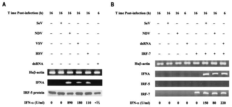
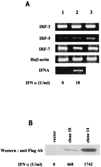
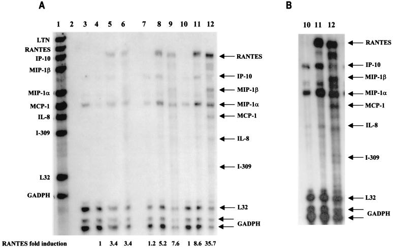
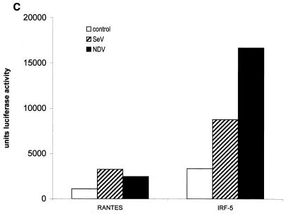
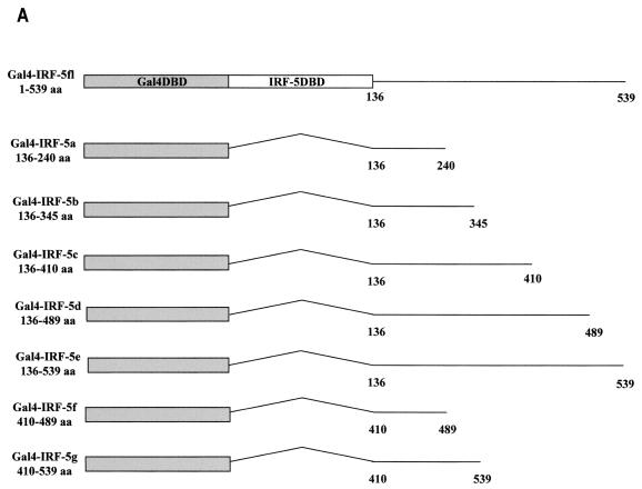
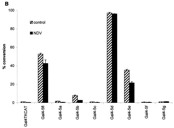
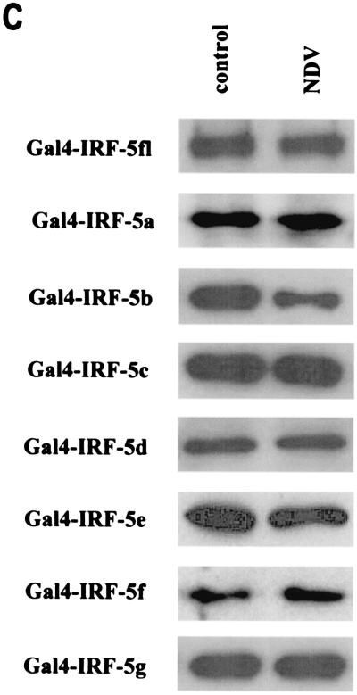
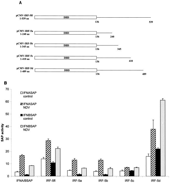
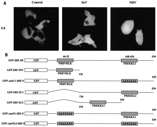
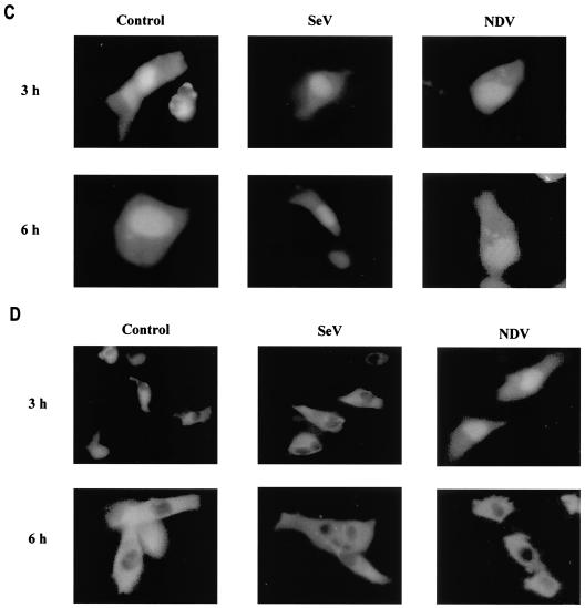
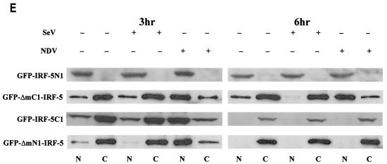
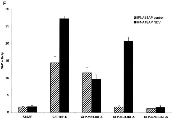
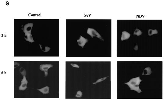
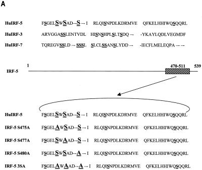
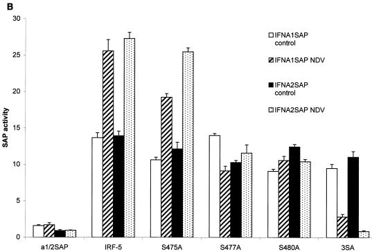
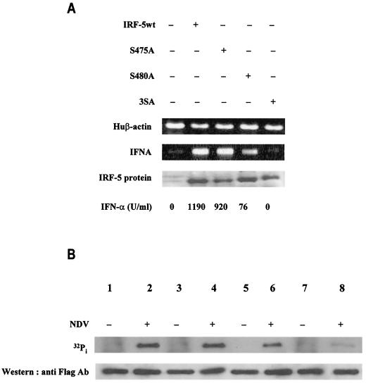
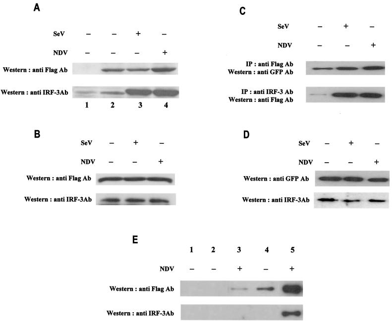
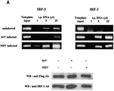
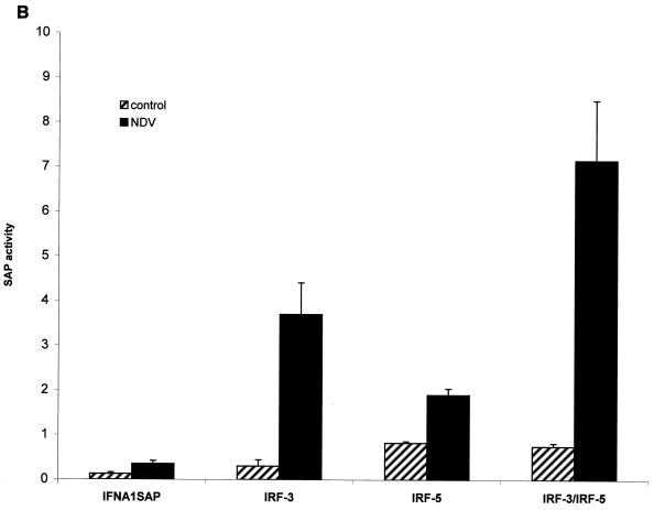
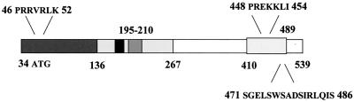
Transcription factors of the interferon regulatory factor (IRF) family have been identified as critical mediators of early inflammatory gene transcription in infected cells. We recently determined that, besides IRF-3 and IRF-7, IRF-5 serves as a direct transducer of virus-mediated signaling. In contrast to that mediated by the other two IRFs, IRF-5-mediated activation is virus specific. We show that, in addition to Newcastle disease virus (NDV) infection, vesicular stomatitis virus (VSV) and herpes simplex virus type 1 (HSV-1) infection activates IRF-5, leading to the induction of IFNA gene subtypes that are distinct from subtypes induced by NDV. The IRF-5-mediated stimulation of inflammatory genes is not limited to IFNA since in BJAB/IRF-5-expressing cells IRF-5 stimulates transcription of RANTES, macrophage inflammatory protein 1 beta, monocyte chemotactic protein 1, interleukin-8, and I-309 genes in a virus-specific manner. By transient- transfection assay, we identified constitutive-activation (amino acids [aa] 410 to 489) and autoinhibitory (aa 490 to 539) domains in the IRF-5 polypeptide. We identified functional nuclear localization signals (NLS) in the amino and carboxyl termini of IRF-5 and showed that both of these NLS are sufficient for nuclear translocation and retention in infected cells. Furthermore, we demonstrated that serine residues 477 and 480 play critical roles in the response to NDV infection. Mutation of these residues from serine to alanine dramatically decreased phosphorylation and resulted in a substantial loss of IRF-5 transactivation in infected cells. Thus, this study defines the regulatory phosphorylation sites that control the activity of IRF-5 in NDV-infected cells and provides further insight into the structure and function of IRF-5. It also shows that the range of IRF-5 immunoregulatory target genes includes members of the cytokine and chemokine superfamilies.


Figures

FIG. 1.
FIG. 1.
Activation of IRF-5 and endogenous IFNA genes by different inducers. (A) Reconstitution of endogenous IFNA gene expression in 2fTGH/IRF-5 cells infected with NDV, VSV, and HSV but not Sendai virus or dsRNA. IFNA cDNAs were amplified using primers corresponding to the conserved regions of all human IFNA genes (61). Human (Hu) β-actin-amplified fragments are shown at the bottom as a control for RNA levels. Levels of IRF-5 in cell lysates were determined by immunoblotting with an anti-Flag antibody, and biologically active IFNA was detected by an antiviral assay using bovine tracheal cells and VSV as the challenging virus (12). (B) 2fTGH cells expressing ectopic IRF-7 induce IFNA gene expression after infection with Sendai virus or NDV and after treatment with dsRNA. 2fTGH cells were left untransfected (left four lanes) or were transfected with IRF-7 expression plasmid (right four lanes) and were uninfected or were infected with Sendai virus or NDV for 16 h or were coincubated with dsRNA and cycloheximide for 6 h. RT-PCR for β-actin, IFNA, IRF-5, and IRF-7 amplification was performed as described in Materials and Methods.
FIG. 2.
FIG. 2.
Activation of endogenous IRFs and IFNA genes in infected BJAB cells. (A) The relative levels of IRF-3, IRF-5, IRF-7, and IFNA mRNA were determined by RT-PCR analysis of BJAB cells (lane 1), NDV-infected BJAB cells at 16 h postinfection (lane 2), and BJAB cells treated with IFN for 8 h (lane 3). Individual cDNAs were amplified by using primers specific for the IRFs or primers corresponding to the conserved regions of all human IFNA genes (4, 7, 61). Levels of biologically active IFN-α synthesized in these cells were determined by antiviral assay and are shown at the bottom. Huβ-actin, human β-actin. (B) The levels of Flag-tagged IRF-5 expressed in stably transfected, NDV-infected BJAB clones 10 and 14 were determined 16 h postinfection by immunoblotting with an anti-Flag antibody. Left lane, empty vector control, pCMVsport; middle lane, low-expressing IRF-5 clone 10; right lane, high-expressing IRF-5 clone 14. The levels of biologically active IFN-α in the medium of infected cells, shown at the bottom, were determined by an antiviral assay (12).
FIG. 3.
FIG. 3.
IRF-5 induces multiple cytokines and chemokines in infected BJAB cells. (A) IRF-5 induces expression of multiple chemokines in infected BJAB cell lines expressing either low levels of Flag-tagged IRF-5 (clone 10) or high levels of Flag-tagged IRF-5 (clone 14). Total RNA was isolated from BJAB/IRF-5 cells at 6 h postinfection and analyzed by RPA using the hCK-5 kit (Pharmingen) as described in Materials and Methods. Lane 1, hCK-5 probe template; lane 2, tRNA control; lane 3, human RNA control; lane 4, uninfected BJAB control cells; lane 5, Sendai virus-infected BJAB control cells; lane 6, NDV-infected BJAB control cells; lane 7, uninfected BJAB/IRF-5 clone 10; lane 8, Sendai virus-infected BJAB/IRF-5 clone 10; lane 9, NDV-infected BJAB/IRF-5 clone 10; lane 10, uninfected BJAB/IRF-5 clone 14; lane 11, Sendai virus-infected BJAB/IRF-5 clone 14; lane 12, NDV-infected BJAB/IRF-5 clone 14. Control cells are BJAB cells transfected with an empty vector. Fold induction of RANTES mRNA expression was normalized to the level of glyceraldehyde-3-phosphate dehydrogenase mRNA expression. The data are representative of two independent experiments. (B) Long exposure of RNA samples analyzed in lanes 10 to 12. (C) Activation of the RANTES luciferase reporter in infected cells expressing IRF-5. HeLa cells were left uninfected or were infected with Sendai virus or NDV for 16 h, and luciferase activity was measured as described in Materials and Methods.
FIG. 3.
FIG. 3.
IRF-5 induces multiple cytokines and chemokines in infected BJAB cells. (A) IRF-5 induces expression of multiple chemokines in infected BJAB cell lines expressing either low levels of Flag-tagged IRF-5 (clone 10) or high levels of Flag-tagged IRF-5 (clone 14). Total RNA was isolated from BJAB/IRF-5 cells at 6 h postinfection and analyzed by RPA using the hCK-5 kit (Pharmingen) as described in Materials and Methods. Lane 1, hCK-5 probe template; lane 2, tRNA control; lane 3, human RNA control; lane 4, uninfected BJAB control cells; lane 5, Sendai virus-infected BJAB control cells; lane 6, NDV-infected BJAB control cells; lane 7, uninfected BJAB/IRF-5 clone 10; lane 8, Sendai virus-infected BJAB/IRF-5 clone 10; lane 9, NDV-infected BJAB/IRF-5 clone 10; lane 10, uninfected BJAB/IRF-5 clone 14; lane 11, Sendai virus-infected BJAB/IRF-5 clone 14; lane 12, NDV-infected BJAB/IRF-5 clone 14. Control cells are BJAB cells transfected with an empty vector. Fold induction of RANTES mRNA expression was normalized to the level of glyceraldehyde-3-phosphate dehydrogenase mRNA expression. The data are representative of two independent experiments. (B) Long exposure of RNA samples analyzed in lanes 10 to 12. (C) Activation of the RANTES luciferase reporter in infected cells expressing IRF-5. HeLa cells were left uninfected or were infected with Sendai virus or NDV for 16 h, and luciferase activity was measured as described in Materials and Methods.
FIG. 4.
FIG. 4.
Analysis of intrinsic transactivation potentials of various IRF-5 regions fused to the Gal4 DBD. (A) Schematic representation of seven IRF-5 deletion mutants. Straight lines and angled lines indicate included and excluded sequences, respectively. The Gal4 DBD and IRF-5 DBD are indicated. (B) Gal4 TKCAT reporter transient transfection assay. HeLa cells were cotransfected with the indicated plasmid expressing Gal4-IRF-5 fusion proteins (2.5 μg), Gal4 TKCAT reporter plasmid (2.5 μg), and the β-galactosidase plasmid (0.2 μg). Cells were uninfected or infected with NDV, and CAT activity was measured 48 h after transfection. Percent conversion after normalizing for β-galactosidase activity is expressed. (C) Levels of transfected IRF-5 protein isolated from uninfected HeLa cells or HeLa cells infected with NDV for 16 h were detected by Western blot analysis with the anti-Gal4 (DBD) polyclonal antibody.
FIG. 4.
FIG. 4.
Analysis of intrinsic transactivation potentials of various IRF-5 regions fused to the Gal4 DBD. (A) Schematic representation of seven IRF-5 deletion mutants. Straight lines and angled lines indicate included and excluded sequences, respectively. The Gal4 DBD and IRF-5 DBD are indicated. (B) Gal4 TKCAT reporter transient transfection assay. HeLa cells were cotransfected with the indicated plasmid expressing Gal4-IRF-5 fusion proteins (2.5 μg), Gal4 TKCAT reporter plasmid (2.5 μg), and the β-galactosidase plasmid (0.2 μg). Cells were uninfected or infected with NDV, and CAT activity was measured 48 h after transfection. Percent conversion after normalizing for β-galactosidase activity is expressed. (C) Levels of transfected IRF-5 protein isolated from uninfected HeLa cells or HeLa cells infected with NDV for 16 h were detected by Western blot analysis with the anti-Gal4 (DBD) polyclonal antibody.
FIG. 5.
FIG. 5.
IRF-5 contains an autoinhibitory domain located between aa 489 and 539 as determined by an IFNA SAP reporter assay. (A) Schematic representation of four IRF-5 carboxyl-terminal deletion mutants. (B) Differential activation of IFNA and IFNB SAP promoters by IRF-5 deletion mutants in NDV-infected cells. 2fTGH cells were cotransfected with IRF-5 deletion plasmids (2.5 μg) and either IFNA1 or IFNB SAP reporter plasmids (2.5 μg) together with the β-galactosidase-expressing plasmid (0.2 μg). At 16 h posttransfection, cells were left uninfected or were infected for 16 h with NDV, and SAP activity was measured as previously described (7).
FIG. 6.
FIG. 6.
IRF-5 contains two functional NLSs. (A) NDV infection but not Sendai virus infection leads to the nuclear accumulation of full-length GFP-IRF-5 6 h postinfection. (B) Schematic representation of GFP-IRF-5 fusion proteins containing both amino- and carboxyl-terminal NLSs (GFP-IRF-5fl) and proteins containing either the amino-terminal NLS (GFP-IRF-5 N1 and GFP-ΔmC1-IRF-5), the carboxyl-terminal NLS (GFP-IRF-5 C1, GFP-IRF-5 C2, and GFP-ΔmN1-IRF-5), or both NLSs mutated (GFP-ΔmNLS-IRF-5). The individual NLS sequences areindicated. (C) Nuclear translocation of GFP-IRF-5 N1 is independent of virus infection. 2fTGH cells were transfected with GFP-IRF-5 N1, and subcellular localization was determined 3 and 6 h after virus infection by fluorescence microscopy as described in Materials and Methods. (D) GFP-IRF-5 C1 is responsive to viral infection yet is not retained in the nucleus. Subcellular localization was determined as described above. (E) The relative levels of GFP-IRF-5 N1, GFP-ΔmC1-IRF-5, GFP-IRF-5 C1, and GFP-ΔmN1-IRF-5 in cytoplasmic (C) and nuclear (N) extracts of uninfected and virus-infected cells were determined by immunoblotting with anti-Flag antibodies to detect IRF-5 localization. (F) Differential activation of the IFNA1 SAP reporter by IRF-5 NLS mutants. 2fTGH cells were cotransfected with IRF-5 expression plasmids (2.5 μg) and an IFNA1 SAP reporter plasmid (2.5 μg) together with the β-galactosidase-expressing plasmid (0.2 μg). At 16 h posttransfection, cells were left uninfected or were infected for 16 h with NDV, and SAP activity was measured as previously described (7). (G) Subcellular localization of GFP-ΔmNLS-IRF-5. The IRF-5 amino- and carboxyl-terminal NLSs are responsible for IRF-5 translocation to the nucleus.
FIG. 6.
FIG. 6.
IRF-5 contains two functional NLSs. (A) NDV infection but not Sendai virus infection leads to the nuclear accumulation of full-length GFP-IRF-5 6 h postinfection. (B) Schematic representation of GFP-IRF-5 fusion proteins containing both amino- and carboxyl-terminal NLSs (GFP-IRF-5fl) and proteins containing either the amino-terminal NLS (GFP-IRF-5 N1 and GFP-ΔmC1-IRF-5), the carboxyl-terminal NLS (GFP-IRF-5 C1, GFP-IRF-5 C2, and GFP-ΔmN1-IRF-5), or both NLSs mutated (GFP-ΔmNLS-IRF-5). The individual NLS sequences areindicated. (C) Nuclear translocation of GFP-IRF-5 N1 is independent of virus infection. 2fTGH cells were transfected with GFP-IRF-5 N1, and subcellular localization was determined 3 and 6 h after virus infection by fluorescence microscopy as described in Materials and Methods. (D) GFP-IRF-5 C1 is responsive to viral infection yet is not retained in the nucleus. Subcellular localization was determined as described above. (E) The relative levels of GFP-IRF-5 N1, GFP-ΔmC1-IRF-5, GFP-IRF-5 C1, and GFP-ΔmN1-IRF-5 in cytoplasmic (C) and nuclear (N) extracts of uninfected and virus-infected cells were determined by immunoblotting with anti-Flag antibodies to detect IRF-5 localization. (F) Differential activation of the IFNA1 SAP reporter by IRF-5 NLS mutants. 2fTGH cells were cotransfected with IRF-5 expression plasmids (2.5 μg) and an IFNA1 SAP reporter plasmid (2.5 μg) together with the β-galactosidase-expressing plasmid (0.2 μg). At 16 h posttransfection, cells were left uninfected or were infected for 16 h with NDV, and SAP activity was measured as previously described (7). (G) Subcellular localization of GFP-ΔmNLS-IRF-5. The IRF-5 amino- and carboxyl-terminal NLSs are responsible for IRF-5 translocation to the nucleus.
FIG. 7.
FIG. 7.
Critical role of IRF-5 serine residues 477 and 480 in NDV-induced transactivation. (A) Carboxyl-terminal amino acid homology regions of IRF-5, IRF-3, and IRF-7. Potential serine phosphorylation sites of IRF-5 and serines phosphorylated in IRF-3 and IRF-7 are underlined. The amino acids targeted for alanine substitution are shown in large letters. Hu, human. (B) Expression plasmids (2.5 μg) encoding wt IRF-5 and IRF-5 with point mutations (S475A, S477A, S480A, and 3SA) were cotransfected with IFNA1 or IFNA2 SAP promoters (2.5 μg) into 2fTGH cells. Cells were left uninfected or were infected with NDV for 16 h, and SAP activity was measured as previously described (7).
FIG. 7.
FIG. 7.
Critical role of IRF-5 serine residues 477 and 480 in NDV-induced transactivation. (A) Carboxyl-terminal amino acid homology regions of IRF-5, IRF-3, and IRF-7. Potential serine phosphorylation sites of IRF-5 and serines phosphorylated in IRF-3 and IRF-7 are underlined. The amino acids targeted for alanine substitution are shown in large letters. Hu, human. (B) Expression plasmids (2.5 μg) encoding wt IRF-5 and IRF-5 with point mutations (S475A, S477A, S480A, and 3SA) were cotransfected with IFNA1 or IFNA2 SAP promoters (2.5 μg) into 2fTGH cells. Cells were left uninfected or were infected with NDV for 16 h, and SAP activity was measured as previously described (7).
FIG. 8.
FIG. 8.
Serine-477 and -480 are phosphorylated in NDV-infected cells and are essential for the IRF-5-mediated activation of IFNA genes. (A) Analysis of IFNA transcripts in 2fTGH cells transfected with wt IRF-5 or point mutant IRF-5 S475A, S480A, or 3SA and infected with NDV. IFNA cDNAs were amplified by PCR with primers corresponding to the regions of IFNA genes that are conserved in all IFNA subtypes (61). The nuclear levels of IRF-5 and mutants were determined by immunoblotting with an anti-Flag antibody. The levels of biologically active IFN-α synthesized in these cells as determined by an antiviral assay are shown at the bottom. (B) Specific phosphorylation of IRF-5 point mutants by NDV infection. 2fTGH cells were transfected with wt IRF-5 (lanes 1 and 2) or IRF-5 point mutants (lanes 3 and 4, S475A; lanes 5 and 6, S480A; lanes 7 and 8, 3SA) and left uninfected (−) or infected (+) with NDV. Cells were incubated for 6 h in media containing [32P]orthophosphate, and then the 32P-labeled IRF-5 was precipitated from cell lysates with an anti-Flag antibody and proteins were separated by electrophoresis in SDS-7% polyacrylamide gel, dried, and then exposed to a PhosphorImager screen (Molecular Dynamics, Inc., Sunnyvale, Calif.). Top, levels of radiolabeled IRF-5; bottom, levels of IRF-5 protein in cell lysates detected by immunoblotting with an anti-Flag antibody.
FIG. 9.
FIG. 9.
Enhanced dimerization of phosphorylated IRF-5 in infected cells. (A) 2fTGH/IRF-5 cells were left uninfected or were infected with Sendai virus or NDV for 6 h. Dimerization of IRF-5 was analyzed by GST pull-down assay (10). Whole-cell extract (250 μg) was applied to GST-agarose beads (lane 1) or GST-tagged IRF-5-agarose beads (lanes 2 to 4), and specifically bound proteins were detected by Western blotting with an anti-Flag antibody (Ab) (top) or an anti-IRF-3 polyclonal antibody (bottom) as described in Materials and Methods. (B) Expression of Flag-tagged IRF-5 and endogenous IRF-3 in the cell lysates analyzed in panel A. Proteins detected represent 8% of input onto GST columns. (C) Detection of IRF-5 dimerization in infected cells. 2fTGH/IRF-5 cells were transfected with GFP-IRF-5 and then infected with Sendai virus or NDV for 6 h. Whole-cell extracts (250 μg) were immunoprecipitated (IP) with either an anti-Flag antibody (top) or an anti-IRF-3 polyclonal antibody (bottom) as described in Materials and Methods. The immunoprecipitated complexes were separated on SDS-7% PAGE gels and subsequently probed with an anti-GFP polyclonal antibody or an anti-Flag antibody to detect IRF-5. (D) Expression levels of endogenous IRF-5 and IRF-3 in the cell lysates of 2fTGH/IRF-5 cells analyzed in panel C. IRF-5 and IRF-3 were detected with an anti-GFP antibody or an anti-IRF-3 antibody, respectively. (E) Formation of the IRF-3/IRF-5 heterodimer is mediated through the IRF-5 carboxyl terminus. Whole-cell lysate (250 μg) from 2fTGH/IRF-5 uninfected or NDV-infected cells, as shown in panel B, was used for mapping the IRF-5 interaction domain. Cell lysates were applied to GST-agarose beads (lane 1) or IRF-5-agarose beads where IRF-5 was tagged with GST at the amino terminus (lanes 2 and 3) or the carboxyl terminus (lanes 4 and 5). Specifically bound proteins were detected by Western blotting with anti-Flag antibodies (top) or anti-IRF-3 antibodies (bottom), as described in Materials and Methods.
FIG. 10.
FIG. 10.
Binding of IRF-5 and IRF-3 to IFNA promoters. (A) In vivo binding of IRF-5 and IRF-3 to the endogenous IFNA promoter as analyzed by the chromatin immunoprecipitation assay. 2fTGH/IRF-5 cells were infected with Sendai virus or NDV or were left uninfected. Cellular DNA and proteins were cross-linked and subjected to the chromatin immunoprecipitation assay as described in Materials and Methods. The immunoprecipitations were performed with either anti-Flag antibody (Ab) or anti-IRF-3 polyclonal antibody to detect IRF-5 and IRF-3 binding, respectively. DNA recovered from chromatin immunoprecipitation by heating was amplified by using universal primers specific for endogenous IFNA genes (61). Template input, amplification of the endogenous IFNA promoter region from DNA-protein complexes before immunoprecipitation. Immunoprecipitated (i.p.) DNA was resuspended in 60 μl of Tris-EDTA. Serial dilutions (1, 5, or 25 μl) were used as templates for PCR amplification to show that the response was in the linear range. Levels of IRF-5 (anti-Flag Ab) and IRF-3 protein in cell lysates as detected by Western blotting are shown. (B) Cooperation between IRF-3 and IRF-5 binding to IFNA1 VRE enhances IRF-5-induced IFNA1 expression. 2fTGH cells were cotransfected with IFNA1 SAP (1 μg) and IRF-3 (2 μg) or IRF-5 (2 μg) or IRF-3 (2 μg) and IRF-5 (2 μg). All transfections were performed with equal amounts of total DNA (5 μg); pUC19 was used as filler DNA. At 16 h posttransfection, cells were left uninfected or were infected with NDV for an additional 16 h, and SAP activity was measured as described in Materials and Methods. SAP activity is expressed after normalizing for β-galactosidase expression.
FIG. 10.
FIG. 10.
Binding of IRF-5 and IRF-3 to IFNA promoters. (A) In vivo binding of IRF-5 and IRF-3 to the endogenous IFNA promoter as analyzed by the chromatin immunoprecipitation assay. 2fTGH/IRF-5 cells were infected with Sendai virus or NDV or were left uninfected. Cellular DNA and proteins were cross-linked and subjected to the chromatin immunoprecipitation assay as described in Materials and Methods. The immunoprecipitations were performed with either anti-Flag antibody (Ab) or anti-IRF-3 polyclonal antibody to detect IRF-5 and IRF-3 binding, respectively. DNA recovered from chromatin immunoprecipitation by heating was amplified by using universal primers specific for endogenous IFNA genes (61). Template input, amplification of the endogenous IFNA promoter region from DNA-protein complexes before immunoprecipitation. Immunoprecipitated (i.p.) DNA was resuspended in 60 μl of Tris-EDTA. Serial dilutions (1, 5, or 25 μl) were used as templates for PCR amplification to show that the response was in the linear range. Levels of IRF-5 (anti-Flag Ab) and IRF-3 protein in cell lysates as detected by Western blotting are shown. (B) Cooperation between IRF-3 and IRF-5 binding to IFNA1 VRE enhances IRF-5-induced IFNA1 expression. 2fTGH cells were cotransfected with IFNA1 SAP (1 μg) and IRF-3 (2 μg) or IRF-5 (2 μg) or IRF-3 (2 μg) and IRF-5 (2 μg). All transfections were performed with equal amounts of total DNA (5 μg); pUC19 was used as filler DNA. At 16 h posttransfection, cells were left uninfected or were infected with NDV for an additional 16 h, and SAP activity was measured as described in Materials and Methods. SAP activity is expressed after normalizing for β-galactosidase expression.
FIG. 11.
FIG. 11.
Schematic representation of IRF-5. Structural and functional domains involved in posttranscriptional modification, subcellular translocation, and interaction with other IRF proteins are shown. aa 1 through 728 constitute the full-length IRF-5 protein. aa 35 through 136 represent the DBD (boxed), containing the tryptophan pentad repeats homologous in all IRF family members. The PEST domain is located between aa 152 and 267, where the internal deletion occurs (aa 195 through 210 [grey box]). A glutamate stretch unique to IRF-5 is located from aa 176 through 183 (black box), and a proline-rich region was identified between aa 189 and 340. IRF-5 also contains a C-terminal protein-interacting domain between aa 268 and 470, which contains the transactivation domain localized at aa 410 through 489 (boxed). Amino acids of the amino- and carboxyl-terminal NLSs are indicated at the top, along with the amino acids of the serine-rich cluster, labeled at the bottom.

Similar articles

Cited by

References

    1. Au, W. C., P. A. Moore, D. W. LaFleur, B. Tombal, and P. M. Pitha. 1998. Characterization of the interferon regulatory factor-7 and its potential role in the transcription activation of interferon A genes. J. Biol. Chem. 273:29210-29217. - PubMed
    1. Au, W.-C., P. A. Moore, W. Lowther, Y.-T. Juang, and P. M. Pitha. 1995. Identification of a member of the interferon regulatory factor family that binds to the interferon-stimulated response element and activates expression of interferon-induced genes. Proc. Natl. Acad. Sci. USA 92:11657-11661. - PMC - PubMed
    1. Au, W. C., and P. M. Pitha. 2001. Recruitment of multiple interferon regulatory factors and histone acetyltransferase to the transcriptionally active interferon a promoters. J. Biol. Chem. 276:41629-41637. - PubMed
    1. Au, W. C., W. S. Yeow, and P. M. Pitha. 2001. Analysis of functional domains of interferon regulatory factor 7 and its association with IRF-3. Virology 280:273-282. - PubMed
    1. Baggiolini, M., B. Dewald, and B. Moser. 1997. Human chemokines: an update. Annu. Rev. Immunol. 15:675-705. - PubMed

Publication types

MeSH terms