. 2002 Aug;22(16):5813-25.
doi: 10.1128/MCB.22.16.5813-5825.2002.

The zinc finger domain of NEMO is selectively required for NF-kappa B activation by UV radiation and topoisomerase inhibitors

Affiliations

The zinc finger domain of NEMO is selectively required for NF-kappa B activation by UV radiation and topoisomerase inhibitors

Tony T Huang et al. Mol Cell Biol. 2002 Aug.

Abstract

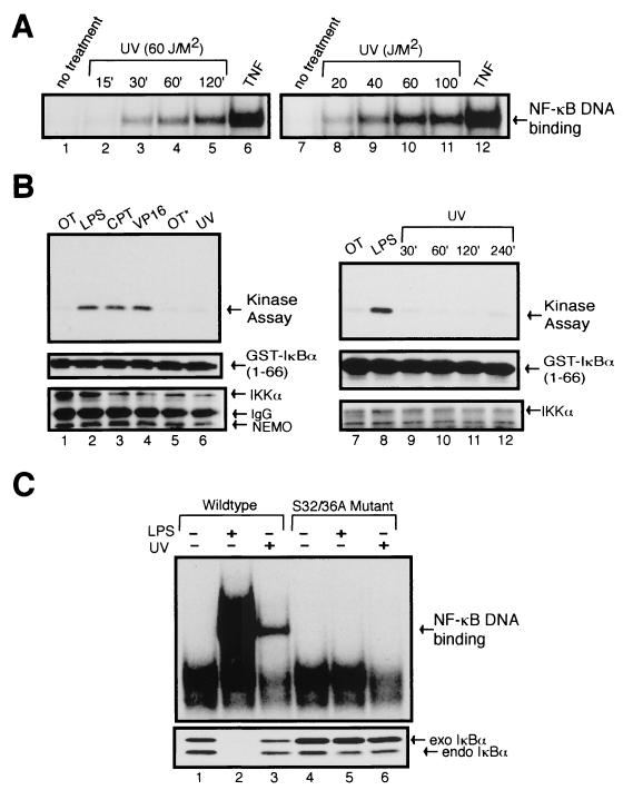
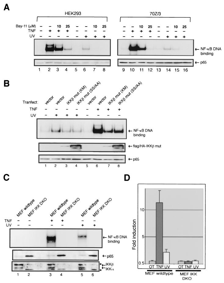
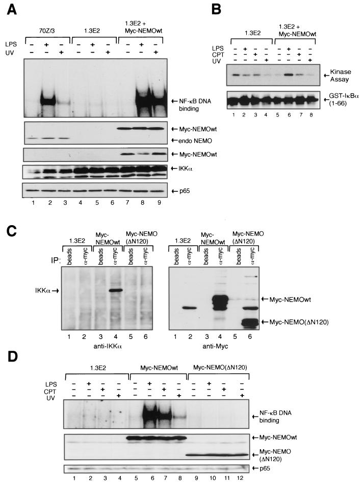
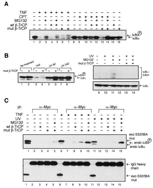
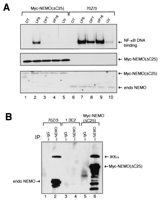
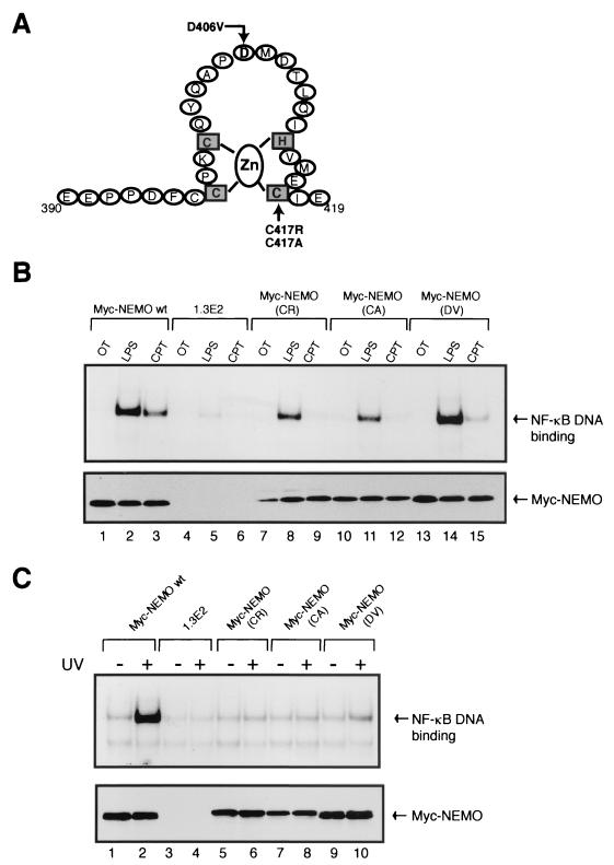
Exposure of mammalian cells to UV radiation was proposed to stimulate the transcription factor NF-kappa B by a unique mechanism. Typically, rapid and strong inducers of NF-kappa B, such as tumor necrosis factor alpha (TNF-alpha) and bacterial lipopolysaccharide (LPS), lead to rapid phosphorylation and proteasomal degradation of its inhibitory protein, I kappa B alpha. In contrast, UV, a relatively slower and weaker inducer of NF-kappa B, was suggested not to require phosphorylation of I kappa B alpha for its targeted degradation by the proteasome. We now provide evidence to account for this peculiar degradation process of I kappa B alpha. The phospho-I kappa B alpha generated by UV is only detectable by expressing a Delta F-box mutant of the ubiquitin ligase beta-TrCP, which serves as a specific substrate trap for serine 32 and 36 phosphorylated I kappa B alpha. In agreement with this finding, we also find that the I kappa B kinase (IKK) phospho-acceptor sites on I kappa B alpha, core components of the IKK signalsome, and IKK catalytic activity are all required for UV signaling. Furthermore, deletion and point mutation analyses reveal that both the amino-terminal IKK-binding and the carboxy-terminal putative zinc finger domains of NEMO (IKK gamma) are critical for UV-induced NF-kappa B activation. Interestingly, the zinc finger domain is also required for NF-kappa B activation by two other slow and weak inducers, camptothecin and etoposide. In contrast, the zinc finger module is largely dispensable for NF-kappa B activation by the rapid and strong inducers LPS and TNF-alpha. Thus, we suggest that the zinc finger domain of NEMO likely represents a point of convergence for signaling pathways initiated by slow and weak NF-kappa B-activating conditions.


Figures

FIG. 1.
FIG. 1.
Stably expressed S32/36A mutant of IκBα inhibits UV-induced NF-κB DNA-binding activity. (A) HeLa cells were either untreated or exposed to UV (60 J/m2) for the indicated time periods (lanes 2 to 5) or exposed to indicated doses of UV for 2 h (lanes 8 to 11) or treated with TNF-α (10 ng/ml) for 15 min for the control. Total cell extracts were prepared and NF-κB DNA-binding activity was determined by EMSA. (B) 70Z/3 cells were either untreated, treated with LPS (10 μg/ml) for 30 min, treated with CPT (10 μM) or VP16 (10 μM), or exposed to UV (60 J/m2) for 2 h (lane 6) or at the indicated time points (lanes 9 to 12). An asterisk indicates the handling of cells similar to that of UV-treated cells (lane 5). Whole-cell lysates were prepared, and IKK activity was measured by the immunecomplex kinase assay (upper panels) (see Materials and Methods). The levels of IKKα, GST-IκBα, and NEMO were determined by immunobloting with antibodies specific to IKKα, GST, and NEMO (lower panels). (C) 70Z/3-CD14 cells were stably transfected by a retroviral infection method with either HA-tagged wild-type or S32/36A mutant mouse IκBα. Stable clones from both wild-type and mutant-expressing pools of cells were picked and analyzed. Total cell extracts from the samples were analyzed by EMSA (upper panel) and Western blotting with anti-IκBα antibody (lower panel). Prior to the treatment conditions, the cells were pretreated with z-VAD (25 μM) for 30 min to prevent caspase-dependent cleavage of IκBα (see Discussion). The wild-type and mutant-expressing cells were left untreated, treated with LPS (1 μg/ml) for 15 min, or exposed to UV (60 J/m2) for 2 h. The exogenous HA-tagged IκBα (exo IκBα) runs slightly higher than the endogenous IκBα (endo IκBα) due to the presence of the epitope tag.
FIG. 2.
FIG. 2.
IKK is required for UV-induced NF-κB activation. (A) HEK293 and 70Z/3 cells were both pretreated with 10 μM (lanes 3, 7, 11, and 15) or 25 μM (lanes 4, 8, 12, and 16) of the IKK inhibitor Bay-11-7082 and then treated with TNF-α (10 ng/ml) for 20 min in HEK293 or LPS (10 μg/ml) in 70Z/3 cells for 30 min as positive controls, or they were exposed to UV (60 J/m2) irradiation for 2 h (70Z/3) or 4 h (HEK293) as indicated. Total cell extracts were made and analyzed by EMSA for NF-κB binding activity and Western blotting for p65 protein for a loading control. (B) HEK293 cells were transiently transfected with either vector control (1.0 μg), FLAG-tagged IKKβ mutant (Lys-to-Met mutation at the putative ATP-binding site; 1.0 μg), or HA-tagged IKKβ activation loop mutant (Ser177/181-to-Ala; 1.0 μg) as indicated. Cells were then either untreated or treated with TNF-α (10 ng/ml) or exposed to UV (60 J/m2) for 4 h as indicated. Total cell extracts were made and analyzed by EMSA for NF-κB binding activity and Western blotting for Flag- and HA-tagged proteins in the same immunoblot with antibodies against both FLAG and HA or for p65 protein expression levels as indicated. The expression levels of IKKβ mut (SS/AA) appear higher possibly due to an efficient HA immunodetection. (C) MEF lines derived from wild-type or IKKα and IKKβ DKO MEFs were either left untreated, treated with TNF-α (10 ng/ml) for15 min, or exposed to UV (60 J/m2) irradiation for 2 h. Total cell extracts were made and analyzed by EMSA for NF-κB binding activity and Western blotting for IKKα, IKKβ and p65 protein expression as indicated. The asterisks indicate nonspecific bands. (D) Wild-type and DKO MEF lines were transiently transfected with an NF-κB-dependent reporter plasmid (3xκB-Luc) and an internal control for transfection efficiency (CMV-β-Gal). At 36 h after transfection, cells were either untreated or treated with TNF-α (10 ng/ml) or exposed to UV (60 J/m2). Cell extracts were analyzed for luciferase and β-Galactosidase activities. Error bars indicate standard deviations. β-Galactosidase activities in DKO MEF were similar to those in wild-type MEF, indicating that the lack of luciferase induction in DKO cells was not due to inefficient transfection of these cells.
FIG. 3.
FIG. 3.
NEMO association with the IKK catalytic core is necessary for UV-induced NF-κB activation. (A) A stably transfected pool of 1.3E2 cells expressing the Myc-tagged wild-type human NEMO, the NEMO-deficient 1.3E2 cells, and the 70Z/3 parental cells were either untreated or treated with LPS (10 μg/ml) for 30 min or exposed to UV (60 J/m2) for 2 h and coanalyzed by EMSA and Western blotting (panels from top to bottom: antibodies against NEMO, c-Myc, IKKα, and p65) as indicated. (B) Myc-NEMO wild-type-expressing pool of 1.3E2 cells and the NEMO-deficient 1.3E2 cells were either untreated or treated with LPS (as described above), CPT (10 μM), or UV for 2 h. Whole-cell lysates were prepared and IKK activity was measured by the immune complex kinase assay (upper panel). Membrane was also immunoblotted with anti-GST antibody for loading control (lower panel). (C) Coimmunoprecipitation studies were done with 1.3E2 cells, Myc-NEMO wild-type-expressing 1.3E2 cells (D10 clone), and Myc-NEMO (ΔN120) cells. Extracts prepared from 107 cells per sample were immunoprecipitated with protein G-Sepharose beads only or with anti-c-Myc antibody overnight at 4°C. Samples were then loaded on an SDS-PAGE gel and examined by immunoblot with anti-IKKα and then anti-c-Myc antibodies as indicated. (D) A pool of 1.3E2 cells stably expressing an N-terminal 120-amino acid-truncated Myc-NEMO (ΔN120) was coanalyzed with 1.3E2 cells and the D10 Myc-NEMO wild-type-expressing 1.3E2 clone. Cells were either untreated or treated with LPS (10 μg/ml) for 30 min, CPT (10 μM) or UV (60 J/m2) for 2 h. Total cell extracts were made and NF-κB binding activity was determined by EMSA. Protein expression levels of c-Myc-tagged proteins and p65 were also examined by Western blotting.
FIG. 4.
FIG. 4.
UV-induced phosphorylation of IκBα is detectable by a ΔF-box mutant of β-TrCP as a specific phospho-protein substrate trap. (A) HEK293 cells were transiently transfected with either empty vector (1.0 μg), Myc-tagged wild-type β-TrCP (1.0 μg), or Myc-tagged ΔF-box β-TrCP (1.0 μg). At 36 h after transfection, cells were either left untreated or were treated with TNF-α (10 ng/ml) for 15 min or CPT (10 μM) for 2 h with or without pretreatment with MG132 (10 μM). Samples were then directly boiled in 2× SDS loading buffer prior to loading them on an SDS-12.5% PAGE gel. pIκBα migrated higher than the basal IκBα in a Western blot assay with an antibody against IκBα. (B) HEK293 cells were transiently transfected with empty vector or the ΔF-box mutant of β-TrCP and either left untreated or treated with TNF-α (as described above) or UV (60 J/m2) at the indicated times or for 4 h in the presence of MG132 (as described above). Cell samples were analyzed as described above. The upper panel of lanes 9 to 14 represents a longer exposure of the upper regions of the immunoblot probed with the anti-IκBα antibody (lower panel). (C) A pool of HEK293 cells stably expressing HA-tagged S32/36A mutant of human IκBα were transiently transfected with either empty vector (1.0 μg), Myc-tagged wild-type β-TrCP (1.0 μg), or Myc-tagged ΔF-box β-TrCP (1.0 μg) as described above and treated as indicated. HEK293 cells were exposed to UV for 4 h for maximal NF-κB activation. A total of 5% of the extracts from untreated or treated samples was directly loaded to distinguish between the exogenous and endogenous IκBα proteins (lanes 1, 6, and 11). Samples were then collected, and cell lysates were made in a buffer containing phosphatase inhibitors (see Materials and Methods). The cell lysates were then subjected to coimmunoprecipitation with the anti-c-Myc antibody and immunoblotted for both endogenous and exogenous IκBα proteins (anti-IκBα antibody, top panel) or exogenous IκBα protein only (anti-HA antibody, bottom panel). Three IκBα forms can be detected (top panel). The lowest band corresponds to the basal endogenous IκBα. The middle band correlates to the endogenous pIκBα. The highest band represents the nondegradable exogenous HA-tagged S32/36A mutant IκBα.
FIG. 5.
FIG. 5.
UV-induced activation of NF-κB selectively requires the C-terminal 25 amino acids of NEMO. (A) 70Z/3, 1.3E2, and Myc-NEMO (ΔC25)-expressing 1.3E2 cells were treated as indicated, and the NF-κB binding activity was analyzed by EMSA (top panel), Myc-NEMO was detected in an immunoblot with an anti-c-Myc antibody (middle panel), and the relative expression levels of both Myc-NEMO (ΔC25) and wild-type endogenous NEMO were compared with a Western blot probed with anti-NEMO antibody (lower panel). (B) Coimmunoprecipitation studies were done with 70Z/3, 1.3E2, and Myc-NEMO (ΔC25)-expressing 1.3E2 cells. A total of 107 cells per sample were lysed and immunoprecipitated with either rabbit immunoglobulin G or anti-NEMO antibodies. The samples were subjected to SDS-PAGE and then immunoblotted with anti-IKKα and -NEMO antibodies.
FIG. 6.
FIG. 6.
NEMO zinc finger point mutations greatly compromise the ability of the cells to activate NF-κB by UV and topoisomerase inhibitors. (A) Diagram depicting point mutations in the Cys2HisCys zinc finger region generated and analyzed in this study. (B) The two known naturally occurring point mutations in the putative zinc finger domain of NEMO (C417R and D406V) were generated as Myc-NEMO (CR) and Myc-NEMO (DV), respectively. Cysteine 417 was also changed to alanine and constructed as Myc-NEMO (CA). The three NEMO point mutants were stably expressed in 1.3E2 cells and analyzed, along with the Myc-NEMO wild-type-expressing 1.3E2 clone D10 and the parental 1.3E2 cells. The five different cell lines were either left untreated or treated with LPS (10 μg/ml) for 30 min or with CPT (10 μM) for 2 h. Total cell extracts were made and analyzed by EMSA for NF-κB binding activity (upper panel) and by Western blotting (anti-c-Myc antibody) for protein expression levels of the Myc-tagged NEMO wild-type and point mutants (lower panel). (C) The five cell lines were also exposed to UV (60 J/m2) for 2 h and analyzed by EMSA (upper panel) and Western blot assay (lower panel) as described above.

References

    1. Bender, K., M. Gottlicher, S. Witeside, H. J. Rahmsdorf, and P. Herrlich. 1998. Sequential DNA damage-independent and -dependent activation of NF-κB by UV. EMBO J. 17:5170-5181. - PMC - PubMed
    1. Cao, Z. D., J. Xiong, M. Takeuchi, T. Kurama, and D. V. Goeddel. 1996. TRAF6 is a signal transducer for interleukin-1. Nature 383:443-446. - PubMed
    1. Chen, Z., J. Hagler, V. J. Palombella, F. Melandri, D. Scherer, D. Ballard, and T. Maniatis. 1995. Signal-induced site-specific phosphorylation targets IκBα to the ubiquitin-proteasome pathway. Genes Dev. 9:1586-1597. - PubMed
    1. Chen, Z. J., L. Parent, and T. Maniatis. 1996. Site-specific phosphorylation of IκBα by a novel ubiquitination-dependent protein kinase activity. Cell 84:853-862. - PubMed
    1. Delhase, M., M. Hayakawa, Y. Chen, and M. Karin. 1999. Positive and negative regulation of IκB kinase activity through IKKβ subunit phosphorylation. Science 284:309-313. - PubMed

Publication types

MeSH terms

Substances