. 2002 Aug;22(16):5859-68.
doi: 10.1128/MCB.22.16.5859-5868.2002.

Interactions between telomerase and primase physically link the telomere and chromosome replication machinery

Affiliations

Interactions between telomerase and primase physically link the telomere and chromosome replication machinery

Saugata Ray et al. Mol Cell Biol. 2002 Aug.

Abstract

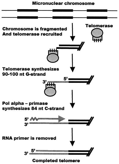
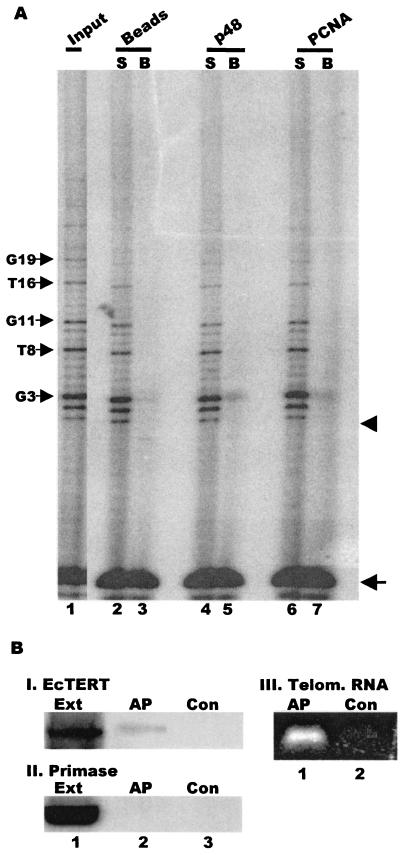
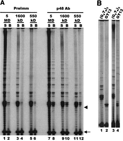
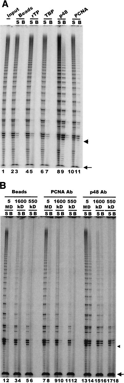
In the ciliate Euplotes crassus, millions of new telomeres are synthesized by telomerase and polymerase alpha-primase during macronuclear development in mated cells. Concomitant with de novo telomere formation, telomerase assembles into higher-order complexes of 550 kDa, 1,600 kDa, and 5 MDa. We show here that telomerase is physically associated with the lagging-strand replication machinery in these complexes. Antibodies against DNA primase precipitated telomerase activity from all three complexes from mated cells but not the 280-kDa telomerase complex from vegetatively growing cells. Moreover, when telomerase was affinity purified, primase copurified with enzyme from mated cells but not with the 280-kDa vegetative complex. Thus, the association of telomerase and primase is developmentally regulated. Intriguingly, PCNA (proliferating cell nuclear antigen) was also found in the 5-MDa complex from mated cells. We therefore speculate that this complex is a complete telomere synthesis machine, while the smaller complexes are assembly intermediates. The physical association of telomerase and primase explains the coordinate regulation of telomeric G- and C-strand synthesis and the efficiency of telomere addition in E. crassus.


Figures

FIG. 1.
FIG. 1.
Cartoon depicting the process of macronuclear development and new telomere synthesis in E. crassus. A 6-nucleotide (nt) 3′ overhang is created by the DNA fragmentation event (24). If C-strand synthesis occurs in several stages, synthesis of the entire G-strand need not necessarily be complete before C-strand synthesis is initiated. The newly synthesized telomeres are trimmed to the mature size at a later stage in macronuclear development (42). Black boxes represent macronucleus-destined sequences.
FIG. 2.
FIG. 2.
Characterization of E. crassus DNA primase. (A) Western blot showing the specificity of the p48 antibody. Lane 1, recombinant, His6-tagged p48; lane 2, macronuclei from vegetatively growing Euplotes cells; lane 3, purified human p48 protein; lane 4, 1.5 × 10⁵ macronuclei from vegetatively growing cells; lane 5, 3.75 × 10⁴ macronuclei from mated cells. The positions of molecular size markers are shown to the left (in kilodaltons). (B) Immunolocalization of primase and E. crassus TERT. Panel I, micro- and macronuclear staining pattern obtained with p48 antibody; panel II, same nuclei stained with Hoechst dye; panel III, staining with E. crassus TERT antibody. The arrows indicate replication bands (RB) and micronuclei (Mic).
FIG. 3.
FIG. 3.
Coimmunoprecipitation of telomerase and primase. (A) Telomerase assay showing activity immunoprecipitated by primase antibody. Protein A beads alone (lane 2 and 3) and beads coated with preimmune IgG (lanes 4 and 5), primase p48 antibody (lanes 6 and 7), or histone H3 variant antibody (lanes 8 and 9) were incubated with nuclear extract and washed, and the beads (B) and unbound supernatant (S) were assayed for telomerase activity. Lane 1 shows the activity in the input extract. The arrowhead marks the position of the input primer. The arrow marks the recovery control that was added to each reaction after the telomerase extension step. (B) Efficiency of primase immunoprecipitation. Primase was immunoprecipitated from extracts made with macronuclei isolated 65 h after mating (lanes 2 and 3) or macronuclei from vegetatively growing cells (lanes 4 and 5) with p48 antibody covalently cross-linked to Sepharose beads. The input and precipitated material were detected with biotinylated p48 antibody and peroxidase-conjugated streptavidin. Lane 1, recombinant p48; lane 2, 5 μl of extract; lane 3, precipitate from 35 μl of extract; lane 4, 10 μl of extract; lane 5, precipitate from 90 μl of extract.
FIG. 4.
FIG. 4.
Affinity purification of telomerase complexes. (A) Schematic of the affinity purification protocol. Following precipitation with NeutrAvidin beads, the affinity-purified material was analyzed by RT-PCR or separated by SDS-polyacrylamide gel electrophoresis, and the composition was examined by Western blotting. (B) Copurification of E. crassus TERT and p48 from extracts made with mated cells. Western blot was probed simultaneously with antibodies to p48 and E. crassus TERT. Lane 1, recombinant E. crassus TERT; lane 2, affinity-purified telomerase complexes. The arrowheads mark p48 and E. crassus TERT. (C) Copurification of telomerase and p48 from 1,600-kDa complexes. The 1,600-kDa complexes were isolated by gel filtration prior to affinity purification. The Western blot was probed with antibody to p48. Lane 1, affinity-purified 1,600-kDa complex; lane 2, recombinant p48. The arrowhead marks full-length p48. Positions of molecular size markers are shown to the left (in kilodaltons).
FIG. 5.
FIG. 5.
Primase is not associated with telomerase from vegetatively growing cells. (A) Immunoprecipitation of telomerase activity. Telomerase assays were performed on the input 280-kDa telomerase complex (lane 1) or the unbound supernatant (S) or bead fraction (B) following incubation with protein A beads alone (lanes 2 and 3) or beads plus p48 antibody (lanes 4 and 5) or PCNA antibody (lanes 6 and 7). The arrows on the left mark the characteristic vegetative telomerase pausing pattern that corresponds to incorporation of the third G and fourth T in the G4T4 repeat. The arrow on the right marks the recovery control, and the arrowhead marks the position of the input primer. (B) Composition of affinity-purified telomerase complexes from vegetatively growing cells. Western blot was probed with antibody to E. crassus TERT (panel I) or p48 (panel II). Lane 1, input vegetative cell extract; lane 2, affinity-purified telomerase; lane 3, control beads incubated with extract but no telomerase-specific oligonucleotide. Panel III, RT-PCR amplification of telomerase RNA from affinity-purified telomerase (lane 1) or control beads incubated with extract but no telomerase-specific oligonucleotide (lane 2).
FIG. 6.
FIG. 6.
Primase is present in all higher-order telomerase complexes. (A) Telomerase assay showing activity immunoprecipitated from partially purified 5-MDa, 1,600-kDa, and 550-kDa complexes. Complexes were precipitated with p48 antibody or preimmune IgG, and unbound supernatant (S) and bead (B) fractions were assayed for each complex with a (G4T4)3 primer. The arrow marks the recovery control, and the arrowhead marks the position of the input primer. (B) Telomerase extension of telomeric and nontelomeric primers. Lanes 1 and 2, purified 550-kDa complex; lanes 3 and 4, unfractionated extract from mated cells. Lanes 1 and 3, telomeric primer (G4T4)3; lanes 2 and 4, nontelomeric primer GT-13.
FIG. 7.
FIG. 7.
PCNA is present in a subset of the telomerase complexes. (A) Interaction with PCNA and telomere-binding proteins. Telomerase assays were performed on the unbound supernatant (S) or the beadfraction (B) following incubation of unfractionated extract from mated cells with antibodies to rTP (lanes 4 and 5), TBP (lanes 6 and 7), p48 (lanes 8 and 9), or PCNA (lanes 10 and 11) or a bead-only control (lanes 2 and 3). Lane 1, activity in input extract. (B) Association of telomerase with PCNA. Immunoprecipitations were performed with purified 5-MDa, 1,600-kDa, and 550-kDa complexes and beads alone (lanes 1 to 6), PCNA (lanes 7 to 12) or p48 antibody (lanes 13 to 18). Arrows mark the recovery control, and arrowheads mark the position of the input primer.

Similar articles

Cited by

References

    1. Adams, A. K., and C. Holm. 1996. Specific DNA replication mutations affect telomere length in Saccharomyces cerevisiae. Mol. Cell. Biol. 16:4614-4620. - PMC - PubMed
    1. Aigner, S., J. Lingner, K. J. Goodrich, C. A. Grosshans, A. Shevchenko, M. Mann, and T. R. Cech. 2000. Euplotes telomerase contains an La motif protein produced by apparent translational frameshifting. EMBO J. 19:6230-6239. - PMC - PubMed
    1. Arezi, B., and R. D. Kuchta. 2000. Eukaryotic DNA primase. Trends Biochem. Sci. 25:572-576. - PubMed
    1. Beattie, T. L., W. Zhou, M. O. Robinson, and L. Harrington. 2001. Functional multimerization of the human telomerase reverse transcriptase. Mol. Cell. Biol. 21:6151-6160. - PMC - PubMed
    1. Bednenko, J., M. Melek, E. C. Greene, and D. E. Shippen. 1997. Developmentally regulated initiation of DNA synthesis by telomerase: evidence for factor-assisted de novo telomere formation. EMBO J. 16:2507-2518. - PMC - PubMed

Publication types

LinkOut - more resources