. 2002 Aug;22(16):5897-911.
doi: 10.1128/MCB.22.16.5897-5911.2002.

Site-directed perturbation of protein kinase C- integrin interaction blocks carcinoma cell chemotaxis

Affiliations

Site-directed perturbation of protein kinase C- integrin interaction blocks carcinoma cell chemotaxis

Maddy Parsons et al. Mol Cell Biol. 2002 Aug.

Abstract

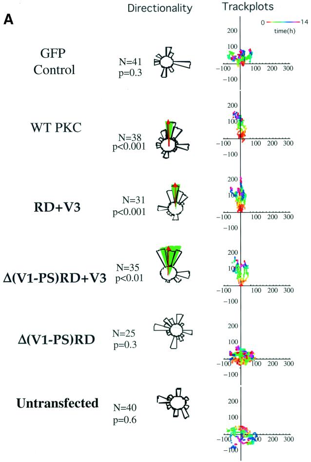
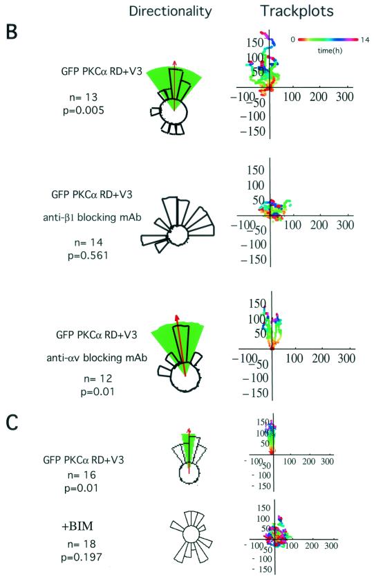
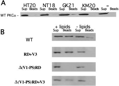
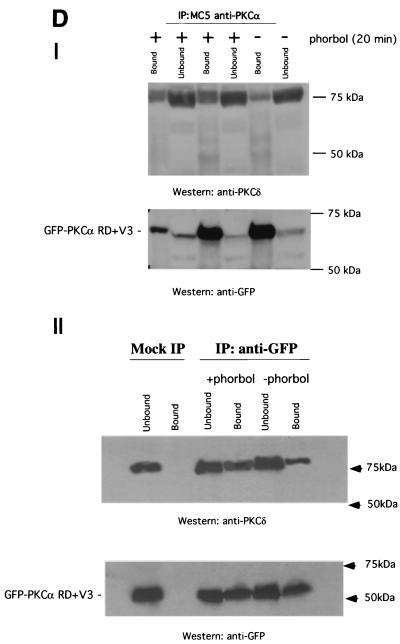
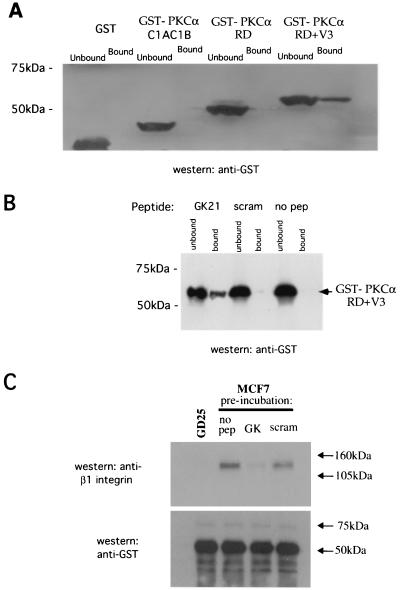
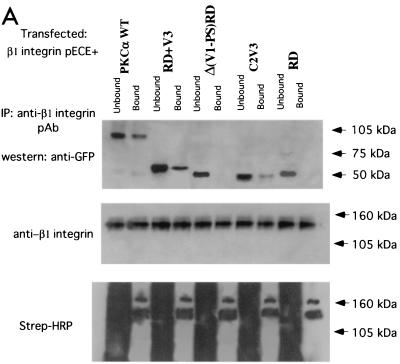
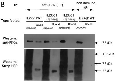
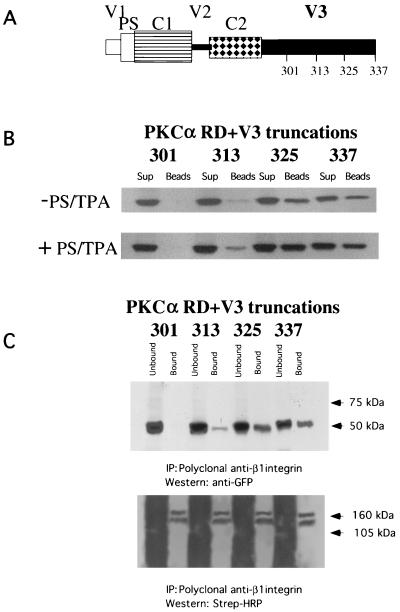
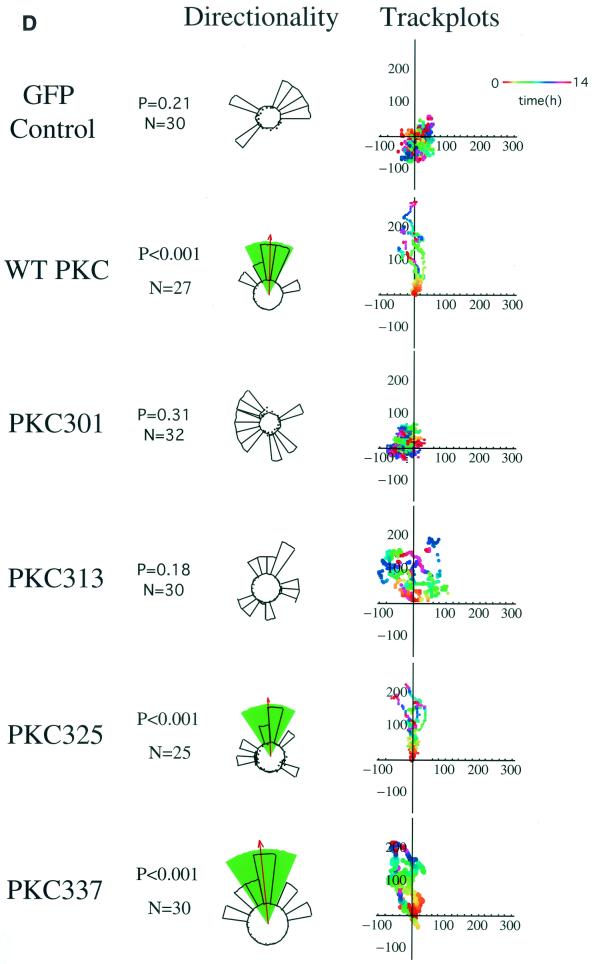
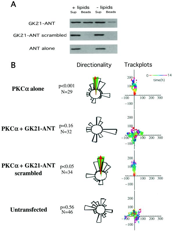
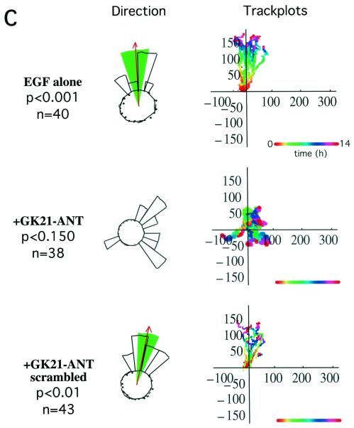
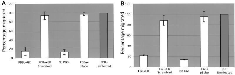
Polarized cell movement is an essential requisite for cancer metastasis; thus, interference with the tumor cell motility machinery would significantly modify its metastatic behavior. Protein kinase C alpha (PKC alpha) has been implicated in the promotion of a migratory cell phenotype. We report that the phorbol ester-induced cell polarization and directional motility in breast carcinoma cells is determined by a 12-amino-acid motif (amino acids 313 to 325) within the PKC alpha V3 hinge domain. This motif is also required for a direct association between PKC alpha and beta 1 integrin. Efficient binding of beta 1 integrin to PKC alpha requires the presence of both NPXY motifs (Cyto-2 and Cyto-3) in the integrin distal cytoplasmic domains. A cell-permeant inhibitor based on the PKC-binding sequence of beta 1 integrin was shown to block both PKC alpha-driven and epidermal growth factor (EGF)-induced chemotaxis. When introduced as a minigene by retroviral transduction into human breast carcinoma cells, this inhibitor caused a striking reduction in chemotaxis towards an EGF gradient. Taken together, these findings identify a direct link between PKC alpha and beta 1 integrin that is critical for directed tumor cell migration. Importantly, our findings outline a new concept as to how carcinoma cell chemotaxis is enhanced and provide a conceptual basis for interfering with tumor cell dissemination.


Figures

FIG. 1.
FIG. 1.
Effects of different GFP-PKCα RD constructs on MCF-7 cell migration in response to a PDBu gradient. (A) MCF-7 cells expressing full-length GFP-PKCα or two RD constructs with the variable region V3 [GFP-PKCα RD+V3 and GFP-PKCα Δ(V1-PS)RD+V3] exhibited significant directional chemotaxis towards a PDBu gradient (1 μM in the outer well of the Dunn chamber) (P < 0.001, P < 0.001, and P < 0.01, respectively). Cells transiently expressing the GFP-PKCα(V1-PS)RD construct, which does not contain the V3 region, or the empty vector, displayed no directionality towards the gradient (P = 0.3). Untransfected cells exhibited no significant directional motility. Analysis of tracked cells from three independent experiments in comparison to vector-alone equivalents was performed using ANOVA. Significant directional chemotaxis towards the PDBu gradient (0°) is represented by a highlighted arc, and the corresponding P value is shown where relevant. The total number of cells tracked (N) is shown. The track plots show all the cell trajectories during the entire time course of each experiment. Each dot represents a cell position at a particular time point which is indicated by the pseudocolor scale (time in hours) beneath each set of cell tracks. The cell track axes are in micrometers. (B) Directional cell motility of cells expressing GFP-PKCα RD+V3 was dependent on β1, but not αV, integrin function. Transiently transfected cells were either left alone or preincubated for 1 h with an anti-β1 (P4C10) or anti-αV integrin (L230) inhibitory MAb (10 μg/ml), before subjected to the chemotaxis assay described above for panel a, in the presence of the corresponding blocking antibody. (C) Directional cell movement of cells expressing GFP-PKCα RD+V3 was inhibited by bisindolylmaleimide (BIM).
FIG. 1.
FIG. 1.
Effects of different GFP-PKCα RD constructs on MCF-7 cell migration in response to a PDBu gradient. (A) MCF-7 cells expressing full-length GFP-PKCα or two RD constructs with the variable region V3 [GFP-PKCα RD+V3 and GFP-PKCα Δ(V1-PS)RD+V3] exhibited significant directional chemotaxis towards a PDBu gradient (1 μM in the outer well of the Dunn chamber) (P < 0.001, P < 0.001, and P < 0.01, respectively). Cells transiently expressing the GFP-PKCα(V1-PS)RD construct, which does not contain the V3 region, or the empty vector, displayed no directionality towards the gradient (P = 0.3). Untransfected cells exhibited no significant directional motility. Analysis of tracked cells from three independent experiments in comparison to vector-alone equivalents was performed using ANOVA. Significant directional chemotaxis towards the PDBu gradient (0°) is represented by a highlighted arc, and the corresponding P value is shown where relevant. The total number of cells tracked (N) is shown. The track plots show all the cell trajectories during the entire time course of each experiment. Each dot represents a cell position at a particular time point which is indicated by the pseudocolor scale (time in hours) beneath each set of cell tracks. The cell track axes are in micrometers. (B) Directional cell motility of cells expressing GFP-PKCα RD+V3 was dependent on β1, but not αV, integrin function. Transiently transfected cells were either left alone or preincubated for 1 h with an anti-β1 (P4C10) or anti-αV integrin (L230) inhibitory MAb (10 μg/ml), before subjected to the chemotaxis assay described above for panel a, in the presence of the corresponding blocking antibody. (C) Directional cell movement of cells expressing GFP-PKCα RD+V3 was inhibited by bisindolylmaleimide (BIM).
FIG. 2.
FIG. 2.
The third variable domain (V3) is required for the lipid-dependent binding of PKCα to a β1 integrin cytoplasmic peptide containing both Cyto-2 and Cyto-3 amino acid motifs. Four N-terminally biotinylated, overlapping peptides based on the cytoplasmic sequence of human β1 integrin were synthesized: KM (aa757-776), KLLMIIHDRREFAKFEKEKM; HT (aa763-782), HDRREFAKFEKEKMNAKWDT; NT, (aa777-794), NAKWDTGENPIYKSAVTT; GK, (aa783-803), GENPIYKSAVTTVVNPKYEGK. (A) In an in vitro pulldown assay (described in Materials and Methods), full-length GFP-PKCα was most efficiently bound to the β1 integrin Cyto-2- and Cyto-3-containing peptide GK. For each sample, all of the peptide-bound GFP-PKCα (Beads) was run alongside one-fifth of the corresponding supernatant containing unbound proteins (Sup). The negative-control lane (−) indicates incubation with streptavidin-coupled agarose beads alone. Immunodetection was performed using an anti-GFP polyclonal antiserum (Clontech). The figure shows a representative result of three experiments. (B) The lipid or activator requirement for WT PKCα and different GFP-tagged PKCα RD constructs to bind to the β1 integrin Cyto domain peptide GK21 in vitro was investigated by preincubating the cell extracts containing the different PKCα constructs with mixed micelles containing PS and TPA (+ lipids), compared with no-lipid or no-activator control (− lipids).
FIG. 3.
FIG. 3.
Structural requirement for PKCα RD in β1 integrin association and its localization in MCF-7 cells. (A) MCF-7 cells were transiently transfected with various GFP-tagged PKCα RD constructs [GFP-Δ(V1-PS)RD+V3, GFP-Δ (V1-PS)RD, and GFP-PKCα RD+V3]. Cells were stimulated with TPA (400 nM) and fixed after 20 min of incubation at 37°C and then either left as controls or stained with MAb 12G10 Fab (+12G10 Fab-Cy3). The fluorescence images from the donor (GFP-PKCα RD) and acceptor (Integrin 12G10 Fab-Cy3) are shown. <τ> is the average of τp and τm, and its pseudocolor scale applies to all four lifetime maps. The FRET efficiency (Eff) pseudocolor plots apply only to the + 12G10 Fab-Cy3 lifetime maps (Eff = 1 − τDAD, where τDA is the lifetime map of the donor in the presence of acceptor and τD is the average lifetime of the donor in the absence of acceptor [average of the mean <τ> of at least four GFP-PKC RD alone control cells]). The pixel count-versus-FRET efficiency graphs to the right summarize the difference between the different RD constructs in their capacities to bind activated β1 integrins [GFP-PKCα RD+V3 (blue trace), GFP-PKCα Δ(V1-PS)RD+V3] (red trace), and GFP-PKCα Δ (V1-PS)RD (green trace)]. Results for GFP-PKCα RD+V3 were similar to those published previously using the same assay (16) and summarized in the pixel count-versus-FRET efficiency graphs for comparison. The cumulative lifetimes of GFP-PKCα donor alone (green) and that measured in the presence of the acceptor fluorophore (red) (n = 5) are plotted on the two-dimensional graphs in the inserts shown to the right. (B) Confocal images of unstimulated, paraformaldehyde-fixed MCF-7 cells coexpressing RFP-PKCα Δ(V1-PS)RD+V3 or RFP-PKCα RD+V3 and GFP-actin. Bars = 10 μm. (C) Still images from supplementary movies 1 and 2 of live MCF-7 cells coexpressing RFP-PKCα RD+V3 (movie 1) or RFP-PKCα Δ(V1-PS)RD+V3 (movie 2) and GFP-actin, simulated with PDBu (1 μM). Frame 001 corresponded to 1 min (movie 1) and 3 min (movie 2) after the addition of PDBu (1 μM) to tissue medium, respectively. Time-lapse series were taken as described in Materials and Methods. During filming, from time to time, the microscope had to be manually adjusted to reestablish the focus, which was less stable with high-power objectives even though the temperature was maintained at 37°C. The white arrows point to RFP-PKCα RD+V3-enriched vesicles that moved in and out of actin-rich cell ruffles (movie 1) and RFP-PKCα Δ(V1-PS)RD+V3-enriched vesicles that trafficked in streams directionally to the leading edge as the cell protruded in response to PDBu stimulation (movie 2). The blue arrows point to the tail of the cell with little or no RFP-PKCα RD+V3 vesicle traffic (movie 1) and a PDBu-induced cell protrusion (movie 2). (D) Panel I shows the coprecipitation of GFP-PKCα RD+V3 with PKCδ, in response to stimulation with phorbol ester (1 μM PDBu) (+). All the bound proteins on the protein G bead (Bound) and one-fifth of the unbound proteins left in the cell extract supernatant after the first centrifugation postprecipitation (Unbound) were loaded. The blot containing the anti-PKCα (MAb MC5) immunoprecipitates was first probed with a rabbit anti-PKCδ polyclonal antibody and then stripped and reprobed with an anti-GFP rabbit serum (Clontech). Duplicates of the +PDBu samples were run. The positions of the molecular mass markers are shown. Panel II is a repeat of the experiment in panel I, with a mock immunoprecipitation (IP) control (i.e., immunoprecipitation with a preimmune serum); immunoprecipitation of GFP-PKCα RD+V3 was performed with a rabbit anti-GFP serum.
FIG. 3.
FIG. 3.
Structural requirement for PKCα RD in β1 integrin association and its localization in MCF-7 cells. (A) MCF-7 cells were transiently transfected with various GFP-tagged PKCα RD constructs [GFP-Δ(V1-PS)RD+V3, GFP-Δ (V1-PS)RD, and GFP-PKCα RD+V3]. Cells were stimulated with TPA (400 nM) and fixed after 20 min of incubation at 37°C and then either left as controls or stained with MAb 12G10 Fab (+12G10 Fab-Cy3). The fluorescence images from the donor (GFP-PKCα RD) and acceptor (Integrin 12G10 Fab-Cy3) are shown. <τ> is the average of τp and τm, and its pseudocolor scale applies to all four lifetime maps. The FRET efficiency (Eff) pseudocolor plots apply only to the + 12G10 Fab-Cy3 lifetime maps (Eff = 1 − τDAD, where τDA is the lifetime map of the donor in the presence of acceptor and τD is the average lifetime of the donor in the absence of acceptor [average of the mean <τ> of at least four GFP-PKC RD alone control cells]). The pixel count-versus-FRET efficiency graphs to the right summarize the difference between the different RD constructs in their capacities to bind activated β1 integrins [GFP-PKCα RD+V3 (blue trace), GFP-PKCα Δ(V1-PS)RD+V3] (red trace), and GFP-PKCα Δ (V1-PS)RD (green trace)]. Results for GFP-PKCα RD+V3 were similar to those published previously using the same assay (16) and summarized in the pixel count-versus-FRET efficiency graphs for comparison. The cumulative lifetimes of GFP-PKCα donor alone (green) and that measured in the presence of the acceptor fluorophore (red) (n = 5) are plotted on the two-dimensional graphs in the inserts shown to the right. (B) Confocal images of unstimulated, paraformaldehyde-fixed MCF-7 cells coexpressing RFP-PKCα Δ(V1-PS)RD+V3 or RFP-PKCα RD+V3 and GFP-actin. Bars = 10 μm. (C) Still images from supplementary movies 1 and 2 of live MCF-7 cells coexpressing RFP-PKCα RD+V3 (movie 1) or RFP-PKCα Δ(V1-PS)RD+V3 (movie 2) and GFP-actin, simulated with PDBu (1 μM). Frame 001 corresponded to 1 min (movie 1) and 3 min (movie 2) after the addition of PDBu (1 μM) to tissue medium, respectively. Time-lapse series were taken as described in Materials and Methods. During filming, from time to time, the microscope had to be manually adjusted to reestablish the focus, which was less stable with high-power objectives even though the temperature was maintained at 37°C. The white arrows point to RFP-PKCα RD+V3-enriched vesicles that moved in and out of actin-rich cell ruffles (movie 1) and RFP-PKCα Δ(V1-PS)RD+V3-enriched vesicles that trafficked in streams directionally to the leading edge as the cell protruded in response to PDBu stimulation (movie 2). The blue arrows point to the tail of the cell with little or no RFP-PKCα RD+V3 vesicle traffic (movie 1) and a PDBu-induced cell protrusion (movie 2). (D) Panel I shows the coprecipitation of GFP-PKCα RD+V3 with PKCδ, in response to stimulation with phorbol ester (1 μM PDBu) (+). All the bound proteins on the protein G bead (Bound) and one-fifth of the unbound proteins left in the cell extract supernatant after the first centrifugation postprecipitation (Unbound) were loaded. The blot containing the anti-PKCα (MAb MC5) immunoprecipitates was first probed with a rabbit anti-PKCδ polyclonal antibody and then stripped and reprobed with an anti-GFP rabbit serum (Clontech). Duplicates of the +PDBu samples were run. The positions of the molecular mass markers are shown. Panel II is a repeat of the experiment in panel I, with a mock immunoprecipitation (IP) control (i.e., immunoprecipitation with a preimmune serum); immunoprecipitation of GFP-PKCα RD+V3 was performed with a rabbit anti-GFP serum.
FIG. 4.
FIG. 4.
Direct association of purified GST-PKCα RD with a β1 integrin cytoplasmic peptide in vitro. (A) In an in vitro pulldown assay (described in Materials and Methods), the binding of GST-PKCα RD+V3, GST-PKCα RD, PKCα C1AC1B (aa32-156), or GST alone to the β1 integrin Cyto-2- and Cyto-3-containing peptide GK was compared. For each sample, all of the peptide-bound GFP-PKCα (Bound) was run alongside one-fifth of the corresponding supernatant containing unbound proteins (Unbound). (B) Binding of GST-PKCα RD+V3 to streptavidin-agarose beads coated with GK, but not its scrambled equivalent (scram) or no-peptide (no pep) control. (C) GST-PKCα RD+V3 on glutathione beads were not treated (no peptide [no pep]) or preincubated with free GK peptide or its scrambled equivalent (scram) (1 mg/ml) for 1 h at 4°C, washed with cell extraction buffer, and then tumbled with cell extracts from either MCF-7 or β1 integrin-null GD25 cells. The bound fraction was then analyzed for β1 integrin and GST-PKCα contents by immunoblotting.
FIG. 5.
FIG. 5.
Binding site mapping by coimmunoprecipitation of human β1 integrin/IL-2R-β1 integrin chimera with PKCα in β1-null GD25 cells. (A) Coprecipitation of β1A integrin (immunoprecipitated with a rabbit polyclonal antiserum against human β1 integrin) with GFP-PKCα WT, GFP-PKCα RD+V3, and GFP-PKCα C2V3, but not with GFP-PKCα Δ(V1-PS)RD, or GFP-PKCα RD, as detected by an anti-GFP antibody. The proteins were from cell surface-biotinylated GD25 cells which were cotransfected with a full-length untagged β1A integrin-pECE and various GFP-tagged PKCα RD constructs. All the bound proteins on the protein G bead (Bound) and one-fifth of the unbound proteins left in the cell extract supernatant after the first centrifugation after precipitation (Unbound) were loaded. The blot containing the anti-integrin immunoprecipitates was stripped and reprobed with an anti-β1 integrin MAb; a similarly prepared duplicate blot was probed with HRP-conjugated streptavidin. Two predominant biotinylated surface proteins were immunoprecipitated using the anti-β1 polyclonal antibody, representing the β1 integrin α/β heterodimer (the lower band comigrates with the β chain detected by the anti-β1 integrin MAb). Positions of the approximate molecular mass markers are shown. (B) Coprecipitation of IL-2R-WT β1A integrin or its truncation constructs (containing the extracellular [EC] and transmembrane [TM] domains of IL-2R and the intracellular [IC] portion of β1A) with endogenous PKCα, as detected by a rabbit polyclonal antiserum against PKCα. The proteins were from GD25 cells transfected with an IL-2R-β1A integrin (aa757-803, WT or aa757-796 or aa757-784) chimera. All the bound proteins on the protein G bead (Bound) and one-fifth of the unbound proteins left in the cell extract supernatant after the first centrifugation postprecipitation (Unbound) were loaded. The blot was stripped and reprobed with HRP-conjugated streptavidin (Strep-HRP).
FIG. 5.
FIG. 5.
Binding site mapping by coimmunoprecipitation of human β1 integrin/IL-2R-β1 integrin chimera with PKCα in β1-null GD25 cells. (A) Coprecipitation of β1A integrin (immunoprecipitated with a rabbit polyclonal antiserum against human β1 integrin) with GFP-PKCα WT, GFP-PKCα RD+V3, and GFP-PKCα C2V3, but not with GFP-PKCα Δ(V1-PS)RD, or GFP-PKCα RD, as detected by an anti-GFP antibody. The proteins were from cell surface-biotinylated GD25 cells which were cotransfected with a full-length untagged β1A integrin-pECE and various GFP-tagged PKCα RD constructs. All the bound proteins on the protein G bead (Bound) and one-fifth of the unbound proteins left in the cell extract supernatant after the first centrifugation after precipitation (Unbound) were loaded. The blot containing the anti-integrin immunoprecipitates was stripped and reprobed with an anti-β1 integrin MAb; a similarly prepared duplicate blot was probed with HRP-conjugated streptavidin. Two predominant biotinylated surface proteins were immunoprecipitated using the anti-β1 polyclonal antibody, representing the β1 integrin α/β heterodimer (the lower band comigrates with the β chain detected by the anti-β1 integrin MAb). Positions of the approximate molecular mass markers are shown. (B) Coprecipitation of IL-2R-WT β1A integrin or its truncation constructs (containing the extracellular [EC] and transmembrane [TM] domains of IL-2R and the intracellular [IC] portion of β1A) with endogenous PKCα, as detected by a rabbit polyclonal antiserum against PKCα. The proteins were from GD25 cells transfected with an IL-2R-β1A integrin (aa757-803, WT or aa757-796 or aa757-784) chimera. All the bound proteins on the protein G bead (Bound) and one-fifth of the unbound proteins left in the cell extract supernatant after the first centrifugation postprecipitation (Unbound) were loaded. The blot was stripped and reprobed with HRP-conjugated streptavidin (Strep-HRP).
FIG. 6.
FIG. 6.
Identification of a 12-amino-acid region within the third variable domain (V3) of PKCα to contain both the lipid-dependent binding site for β1 integrin and molecular determinant for directional cell movement. (A) Schematic representation of various PKCα RD+V3 truncation constructs. (B) Complex formation between various GFP-tagged PKCα V3 domain truncation constructs expressed in MCF-7 cells (in the presence [+] and absence [−] of PS and TPA): 301 (aa1-301), 313 (aa1-313), 325 (aa1-325), and 337 (aa1-337), with the biotinylated GK21 peptide as described in the legend to Fig. 2B. (C) Coprecipitation of β1A integrin (immunoprecipitated [IP] with a rabbit polyclonal antiserum against human β1 integrin) with PKCα 325 (aa1-325) and 337 (aa1-337), as detected by an anti-GFP antibody. The proteins were from cell surface-biotinylated GD25 cells which were cotransfected with a full-length untagged β1A integrin-pECE and the aforementioned GFP-tagged PKCα V3 domain truncations. All the bound proteins on the protein G bead (Bound) and one-fifth of the unbound proteins left in the cell extract supernatant after the first centrifugation after precipitation (Unbound) were loaded. The blot containing the anti-integrin immunoprecipitates was stripped and reprobed with HRP-conjugated streptavidin (Strep-HRP). Two predominant biotinylated surface proteins were immunoprecipitated using the anti-β1 polyclonal antibody, representing the β1 integrin α/β heterodimer, as shown in Fig. 5A. Positions of the approximate molecular mass markers are shown. (D) Effects of the same PKCα V3 domain truncation constructs on directional cell motility towards a PDBu gradient, in comparison with GFP vector control-transfected cells (repeat of the experiment in Fig. 1 using different V3 truncation mutants) (see the legend to Fig. 1A).
FIG. 6.
FIG. 6.
Identification of a 12-amino-acid region within the third variable domain (V3) of PKCα to contain both the lipid-dependent binding site for β1 integrin and molecular determinant for directional cell movement. (A) Schematic representation of various PKCα RD+V3 truncation constructs. (B) Complex formation between various GFP-tagged PKCα V3 domain truncation constructs expressed in MCF-7 cells (in the presence [+] and absence [−] of PS and TPA): 301 (aa1-301), 313 (aa1-313), 325 (aa1-325), and 337 (aa1-337), with the biotinylated GK21 peptide as described in the legend to Fig. 2B. (C) Coprecipitation of β1A integrin (immunoprecipitated [IP] with a rabbit polyclonal antiserum against human β1 integrin) with PKCα 325 (aa1-325) and 337 (aa1-337), as detected by an anti-GFP antibody. The proteins were from cell surface-biotinylated GD25 cells which were cotransfected with a full-length untagged β1A integrin-pECE and the aforementioned GFP-tagged PKCα V3 domain truncations. All the bound proteins on the protein G bead (Bound) and one-fifth of the unbound proteins left in the cell extract supernatant after the first centrifugation after precipitation (Unbound) were loaded. The blot containing the anti-integrin immunoprecipitates was stripped and reprobed with HRP-conjugated streptavidin (Strep-HRP). Two predominant biotinylated surface proteins were immunoprecipitated using the anti-β1 polyclonal antibody, representing the β1 integrin α/β heterodimer, as shown in Fig. 5A. Positions of the approximate molecular mass markers are shown. (D) Effects of the same PKCα V3 domain truncation constructs on directional cell motility towards a PDBu gradient, in comparison with GFP vector control-transfected cells (repeat of the experiment in Fig. 1 using different V3 truncation mutants) (see the legend to Fig. 1A).
FIG. 7.
FIG. 7.
Effect of a cell-permeant form of GK21 on breast carcinoma cell chemotaxis. (A) In the presence of PS and TPA (+ lipids), full-length GFP-PKCα was bound to the GK21 peptide manufactured by chemical synthesis in tandem with the antennapedia third helix (residues 43 to 58) sequence (GK21-ANT), but not its scrambled version (GK21-ANT scrambled) or the antennapedia sequence alone (ANT). For each sample, all of the peptide-bound GFP-PKCα (Beads) was run alongside one-fifth of the corresponding supernatant containing unbound proteins (Sup). (B) MCF-7 cells transiently expressing the full-length GFP-PKCα exhibited significant directional chemotaxis towards a PDBu gradient in a Dunn chamber (P < 0.001). This PKC-driven directional motility was eliminated by pretreating cells with 8 μM of a cell-permeant (antennapedia-tagged), rhodaminated form of GK21 (GK21-ANT), but not its scrambled version. See the legend to Fig. 1A for explanation of track plots. (C) Untransfected MDA-MB-231 cells exhibited significant directional chemotaxis towards an EGF gradient in a Dunn chamber (0.1 μM in the outer well, P < 0.001). Chemotaxis towards EGF was eliminated by pretreating cells with 8 μM of a cell-permeant (antennapedia-tagged), rhodaminated form of GK21 (GK21-ANT), but not its scrambled version.
FIG. 7.
FIG. 7.
Effect of a cell-permeant form of GK21 on breast carcinoma cell chemotaxis. (A) In the presence of PS and TPA (+ lipids), full-length GFP-PKCα was bound to the GK21 peptide manufactured by chemical synthesis in tandem with the antennapedia third helix (residues 43 to 58) sequence (GK21-ANT), but not its scrambled version (GK21-ANT scrambled) or the antennapedia sequence alone (ANT). For each sample, all of the peptide-bound GFP-PKCα (Beads) was run alongside one-fifth of the corresponding supernatant containing unbound proteins (Sup). (B) MCF-7 cells transiently expressing the full-length GFP-PKCα exhibited significant directional chemotaxis towards a PDBu gradient in a Dunn chamber (P < 0.001). This PKC-driven directional motility was eliminated by pretreating cells with 8 μM of a cell-permeant (antennapedia-tagged), rhodaminated form of GK21 (GK21-ANT), but not its scrambled version. See the legend to Fig. 1A for explanation of track plots. (C) Untransfected MDA-MB-231 cells exhibited significant directional chemotaxis towards an EGF gradient in a Dunn chamber (0.1 μM in the outer well, P < 0.001). Chemotaxis towards EGF was eliminated by pretreating cells with 8 μM of a cell-permeant (antennapedia-tagged), rhodaminated form of GK21 (GK21-ANT), but not its scrambled version.
FIG. 8.
FIG. 8.
Retroviral transduction of a minigene insert encoding the GK21 sequence eliminated EGF-induced chemotaxis. MDA-MB231 cells were infected with media from Phoenix packaging cells containing the intact retrovirus encoding the myc epitope-tagged GK21 β1 cytoplasmic sequence, its scrambled equivalent, or the pBabe vector alone. Retroviral transduction efficiency was checked using parallel cultures that were fixed and stained with an anti-myc MAb 9E10. At 48 h postinfection, 231 cells were trypsinized and subjected to an 8-h Transwell chamber migration assay (described in Materials and Methods). Cell harvesting and counting were performed in a blind manner by two individuals, and the subsequent decoding of the samples was carried out by a third investigator. Approximately 10 and 14% of uninfected cells migrated through the filter towards the PDBu (A) and EGF (B) gradients, respectively, over the 8-h time course. The percentage of the cell population that migrated (lower well cell count/upper well cell count) following each treatment was normalized against the mean percentage of uninfected cells that migrated (set at 100%) in the same experiment. Presented are the mean percentages of cells that migrated ± standard deviations from n = 3 wells for each treatment. Results are representative of three independent experiments.
FIG. 9.
FIG. 9.
The effect of introducing retroviral constructs encoding the pBABE vector alone, GK21, or scrambled sequence of GK21 on cell survival was evaluated by a TUNEL assay (Promega). Positive-control cells were incubated with 30 μM genistein for 48 h. Apoptosis was monitored by the amount of TdT-mediated fluorescein-conjugated dUTP uptake, as detected by fluorescence-activated cell sorting analysis. M1 denotes the subset of cells displaying a significant degree of apoptosis above the negative cutoff, which was determined by the amount of fluorescein-conjugated dUTP incorporated in the absence of TdT. Results are representative of two independent experiments.

References

    1. Bailly, M., J. Wyckoff, B. Bouzahzah, R. Hammerman, V. Sylvestre, M. Cammer, R. Pestell, and J. E. Segall. 2000. Epidermal growth factor receptor distribution during chemotactic responses. Mol. Biol. Cell 11:3873-3883. - PMC - PubMed
    1. Bastiaens, P. I. H., and T. M. Jovin. 1998. Fluorescence resonance energy transfer microscopy, p. 136-146. In J. Celis (ed.), Cell biology: a laboratory handbook. Academic Press, New York, N.Y.
    1. Berditchevski, F., G. Bazzoni, and M. E. Hemler. 1995. Specific association of CD63 with the VLA-3 and VLA-6 integrins. J. Biol. Chem. 270:17784-17790. - PubMed
    1. Berrier, A. L., A. M. Mastrangelo, J. Downward, M. Ginsberg, and S. E. LaFlamme. 2000. Activated R-Ras, Rac1, PI 3-kinase and PKCΕ can each restore cell spreading inhibited by isolated integrin β1 cytoplasmic domains. J. Cell Biol. 151:1549-1560. - PMC - PubMed
    1. Bretscher, M. S. 1996. Moving membrane up to the front of migrating cells. Cell 85:465-467. - PubMed

Publication types

MeSH terms