Epiretinal pathology of vitreomacular traction syndrome
- PMID: 12140213
- PMCID: PMC1771255
- DOI: 10.1136/bjo.86.8.902
Epiretinal pathology of vitreomacular traction syndrome
Abstract
Aims: To investigate the ultrastructure of the vitreoretinal interface in patients with vitreomacular traction syndrome.
Methods: 14 patients with vitreomacular traction syndrome underwent standard pars plana vitrectomy. After induction of posterior vitreous detachment, epiretinal tissue and the inner limiting membrane (ILM) of the retina were removed, and processed for transmission electron microscopy.
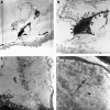
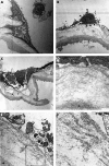
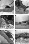
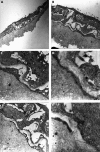
Results: Ultrastructural analysis revealed two basic patterns of vitreoretinal pathology in eyes with vitreomacular traction syndrome. Seven specimens showed mostly single cells or a cellular monolayer covering closely the vitreal side of the ILM, not resulting in a biomicroscopically detectable epiretinal fibrocellular proliferation. The other seven specimens revealed premacular fibrocellular tissue which was separated from the ILM by a layer of native collagen, resembling the clinical features of idiopathic epiretinal membranes. In both groups of eyes, the myofibroblast was the predominant cell type. Fibrous astrocytes and fibrocytes were less frequent. Retinal pigment epithelial cells and macrophages were absent. Deposits of newly formed collagen were present only adjacent to fibrocellular multilayers.
Conclusions: There are two distinct clinicopathological features of vitreomacular traction syndrome which suggest different forms of epiretinal fibrocellular proliferation: (1) epiretinal membranes interposed in native vitreous collagen and (2) single cells or a cellular monolayer proliferating directly on the ILM. The presence of remnants of the cortical vitreous which remain attached to the ILM following posterior vitreous separation may determine the clinicopathological feature of the disease. The predominance of myofibroblasts may help to explain the high prevalence of cystoid macular oedema and progressive vitreomacular traction characteristic for this disorder.
Figures


References
-
- Maumenee AE. Further advances in the study of the macula. Arch Ophthalmol 1967;78:151–65. - PubMed
-
- McDonald HR, Johnson RN, Schatz H. Surgical results in the vitreomacular traction syndrome. Ophthalmology 1994;101:1397–402. - PubMed
-
- Melberg NS, Williams DF, Balles MW, et al. Vitrectomy for vitreomacular traction syndrome with macular detachment. Retina 1995;15:192–7. - PubMed
-
- Smiddy WE, Michels RG, Glaser BM, et al. Vitrectomy for macular traction caused by incomplete vitreous separation. Arch Ophthalmol 1988;106:624–8. - PubMed
-
- Smiddy WE, Michels RG, Green WR. Morphology, pathology, and surgery of idiopathic vitreoretinal macular disorders. A review. Retina 1990;10:288–96. - PubMed
MeSH terms
Substances
LinkOut - more resources
Full Text Sources
Medical
Miscellaneous
