. 2002 Aug 1;30(15):3443-8.
doi: 10.1093/nar/gkf460.

Identification of 5-formyluracil DNA glycosylase activity of human hNTH1 protein

Affiliations

Identification of 5-formyluracil DNA glycosylase activity of human hNTH1 protein

Izumi Miyabe et al. Nucleic Acids Res. .

Abstract

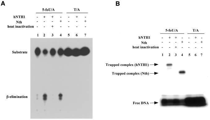
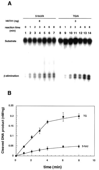
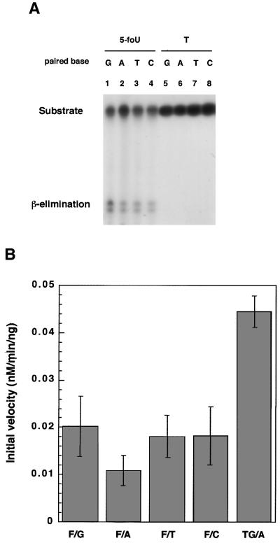
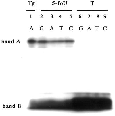
5-formyluracil (5-foU) is a potentially mutagenic lesion of thymine produced in DNA by ionizing radiation and various chemical oxidants. The elucidation of repair mechanisms for 5-foU will yield important insights into the biological consequences of the lesion. Recently, we reported that 5-foU is recognized and removed from DNA by Escherichia coli enzymes Nth (endonuclease III), Nei (endonuclease VIII) and MutM (formamidopyrimidine DNA glycosylase). Human cells have been shown to have enzymatic activities that release 5-foU from X-ray-irradiated DNA, but the molecular identities of these activities are not yet known. In this study, we demonstrate that human hNTH1 (endonuclease III homolog) has a DNA glycosylase/AP lyase activity that recognizes 5-foU in DNA and removes it. hNTH1 cleaved 5-foU-containing duplex oligonucleotides via a beta-elimination reaction. It formed Schiff base intermediates with 5-foU-containing oligonucleotides. Furthermore, hNTH1 cleaved duplex oligonucleotides containing all of the 5-foU/N pairs (N = G, A, T or C). The specific activities of hNTH1 for cleavage of oligonucleotides containing 5-foU and thymine glycol were 0.011 and 0.045 nM/min/ng protein, respectively. These results indicate that hNTH1 has DNA glycosylase activity with the potential to recognize 5-foU in DNA and remove it in human cells.


Figures

Figure 1
Figure 1
Expression and purification of recombinant hNTH1. Proteins were assayed by 12% SDS–PAGE and stained with Coomassie blue. Lane 1, soluble fraction from E.coli induced by 0.1 mM IPTG for 4 h and disrupted by sonication; lane 2, flow-through fraction from glutathione–Sepharose 4B column; lane 3, fraction containing GST–hNTH1 fusion protein eluted with glutathione; lane 4, GST–hNTH1 fusion protein digested by thrombin (5 U/ml); lane 5, purified hNTH1 after elution from a glutathione– Sepharose 4B column. Truncated (lane 6) and full-length (lane 7) hNTH1 were separated by fractionation on a Mono S column. Bands A–C represent GST–hNTH1 fusion, full-length and truncated hNTH1, respectively.
Figure 2
Figure 2
Cleavage activity of hNTH1 on duplex oligonucleotides with or without 5-foU. (A) Cleavage of 5-foU-containing oligonucleotides by purified hNTH1. The duplex oligonucleotide, 5-foU/A (lanes 1–4) or T/A (lanes 5–7), was incubated at 37°C for 20 min without protein (lanes 1 and 5) or with 24 ng hNTH1 (lanes 2 and 6), 25 ng E.coli Nth (lanes 4 and 7) or 24 ng heat-inactivated hNTH1 (lane 3). The inactivation of hNTH1 was performed by heating at 100°C for 10 min. After the reaction was terminated, the products were separated by denaturing 20% PAGE on gels containing 7 M urea. (B) Formation of Schiff base intermediate with 5-foU-containing oligonucleotides by hNTH1. The duplex oligonucleotide substrates were incubated with or without enzyme at 37°C for 5 min in the presence of 50 mM NaBH4. The amounts of hNTH1 and Nth in the reaction mixture were 200 and 100 ng, respectively.
Figure 3
Figure 3
Cleavage of oligonucleotides containing 5-foU and Tg by hNTH1. (A) The duplex oligonucleotides 5-foU/A (lane 1–7) and Tg/A (lane 8–14) were incubated with hNTH1 at 37°C for 0 (lanes 1 and 8), 1 (lanes 2 and 9), 2 (lanes 3 and 10), 3 (lanes 4 and 11), 4 (lanes 5 and 12), 6 (lanes 6 and 13) or 8 (lanes 7 and 14) min. In the reaction mixture, 8 and 3 ng hNTH1 were added for 5-foU and Tg, respectively. The products were separated by denaturing 20% PAGE in gels containing 7 M urea. (B) The intensity of each band was analyzed using NIH Image software. Error bars show the standard deviations.
Figure 4
Figure 4
Effects of the base paired with 5-foU on the cleavage of oligonucleotides by hNTH1. (A) The duplex oligonucleotide substrate, 5-foU/N (lanes 1–4) or T/N (lanes 5–8), was incubated with hNTH1 (8 ng) at 37°C for 3 min. N represents G (lanes 1 and 5), A (lanes 2 and 6), T (lanes 3 and 7) or C (lanes 4 and 8). The products were separated by denaturing 20% PAGE in gels containing 7 M urea. (B) The intensity of the upper band was analyzed using NIH Image software. Error bars show standard deviations.
Figure 5
Figure 5
Trapping assay of purified hNTH1 with NaBH4. The duplex oligonucleotide Tg/A (lane 1), 5-foU/N (lanes 2–5) or T/N (lanes 6–9) was incubated with 100 ng hNTH1 at 37°C for 5 min in the presence of 50 mM NaBH4. N represents G (lanes 2 and 6), A (lanes 3 and 7), T (lanes 4 and 8) or C (lanes 5 and 9).

References

    1. Halliwell B. and Gutteridge,J.M.C. (1990) Role of free radicals and catalytic metal ions in human disease: overview. Methods Enzymol., 186, 1–85. - PubMed
    1. Ames B.N., Shigenaga,M.K. and Hagen,T.M. (1993) Oxidants, antioxidants, and the degenarative diseases of aging. Proc. Natl Acad. Sci. USA, 90, 7915–7922. - PMC - PubMed
    1. Wallace S.S. (1997) Oxidative damage to DNA and its repair. In Scandalios,J.G. (ed.), Oxidative Stress and the Molecular Biology of Antioxidant Defenses. Cold Spring Harbor Laboratory Press, Cold Spring Harbor, NY, pp. 49–81.
    1. Cadet J., Delatour,T., Douki,T., Gasparutto,D., Pouget,J.P., Ravanat,J.L. and Sauvaigo,S. (1999) Hydroxyl radicals and DNA base damage. Mutat. Res., 424, 9–21. - PubMed
    1. Friedberg E.C., Walker,G. and Siede,W. (1995) DNA Repair and Mutagenesis. ASM Press, Washington, DC.

Publication types