. 2002 Aug 6;99(16):10476-81.
doi: 10.1073/pnas.162356199. Epub 2002 Jul 24.

Sonic hedgehog rescues cranial neural crest from cell death induced by ethanol exposure

Affiliations

Sonic hedgehog rescues cranial neural crest from cell death induced by ethanol exposure

Sara C Ahlgren et al. Proc Natl Acad Sci U S A. .

Abstract

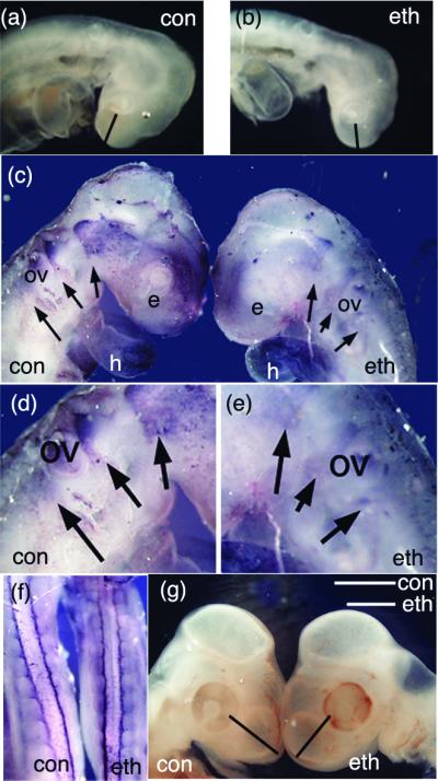
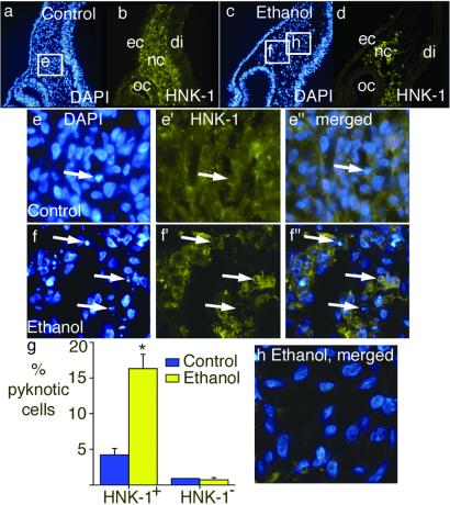
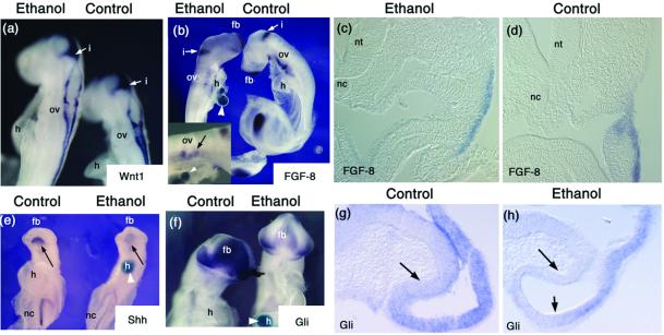
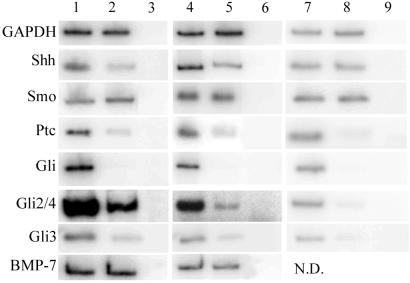
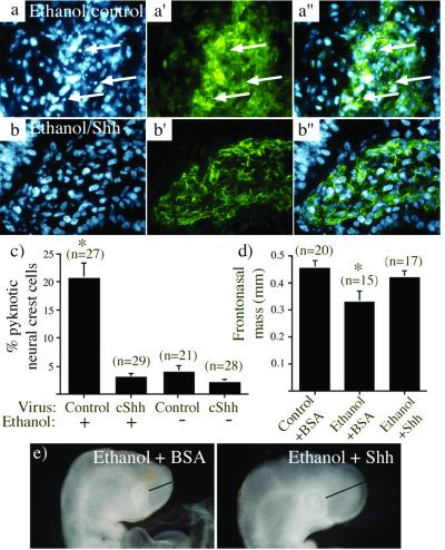
Alcohol is a teratogen that induces a variety of abnormalities including brain and facial defects [Jones, K. & Smith, D. (1973) Lancet 2, 999-1001], with the exact nature of the deficit depending on the time and magnitude of the dose of ethanol to which developing fetuses are exposed. In addition to abnormal facial structures, ethanol-treated embryos exhibit a highly characteristic pattern of cell death. Dying cells are observed in the premigratory and migratory neural crest cells that normally populate most facial structures. The observation that blocking Sonic hedgehog (Shh) signaling results in similar craniofacial abnormalities prompted us to examine whether there was a link between this aspect of fetal alcohol syndrome and loss of Shh. We demonstrate that administration of ethanol to chick embryos results in a dramatic loss of Shh, as well as a loss of transcripts involved in Shh signaling pathways. In contrast, other signaling molecules examined do not demonstrate such dramatic changes. Furthermore, we demonstrate that both the ethanol-induced cranial neural crest cell death and the associated craniofacial growth defect can be rescued by application of Shh. These data suggest that craniofacial anomalies resulting from fetal alcohol exposure are caused at least partially by loss of Shh and subsequent neural crest cell death.


Figures

Fig 1.
Fig 1.
Ethanol treatment results in a reduction in the size of craniofacial tissues. (a and b) Embryos collected approximately 24 h after ethanol treatment (stage 9–10) show distinct differences in craniofacial growth. The measurements for the frontonasal mass are indicated by the bar in each figure; control, 0.42 mm; ethanol, 0.33 mm (see Table 1). (cf) HNK-1 whole-mount analysis of embryos ≈1 day after ethanol treatment demonstrates a reduction in the HNK-1-positive cranial neural crest. Streams of neural crest are indicated with arrows, and the region around the otic vesicle (ov) is enlarged in d and e. Although crest is clearly present in all appropriate regions, the intensity of staining appears reduced in the whole-mount. HNK-1 staining of non-neural tissue, including the heart (h) appears unchanged. (f) The trunk region of ethanol-treated embryos did not display any neural crest abnormalities when stained with HNK-1. (g) Ethanol-treated (stage 9–10) embryos collected 3 days after treatment show distinct size differences of the frontonasal mass (Table 1).
Fig 2.
Fig 2.
There is increased cranial neural crest cell death after ethanol treatment. Embryos were treated with ethanol at stage 9–10, collected 1 day later, fixed, sectioned, and the sections stained for the neural crest marker HNK-1 (in green) and the nuclear marker DAPI (in blue). (a and b) A section taken from the optic cup (oc) region of a control embryo contains HNK-1-positive neural crest cells (nc). (c and d) A section taken from the same region of an ethanol-treated embryo also contains HNK-1-positive neural crest cells. ec, ectoderm; di, diencephalon. (ee′′). Magnified views of the area outlined in a. A single pyknotic nucleus is present (arrow). This nucleus has an HNK-1-positive membrane (e′ and e′′). (ff′′). Magnified views of the area outlined in c. Arrows point to three examples of pyknotic nuclei in f, all of which have HNK-1-positive membranes (f′). (g) Analysis of the pyknotic cells demonstrates that exposure to ethanol resulted in a dramatic increase in the percentage of the cranial neural crest cells (HNK-1-positive) that appear pyknotic, whereas no change in the survival of the non-neural crest mesenchyme (HNK-1-negative) was observed. HNK-1-positive: control n = 44; ethanol n = 48. *, Unpaired t test, P < 0.0001. HNK-1-negative: control n = 44; ethanol n = 39, P not significant. (h) Merged image of DAPI and HNK-1 from the region marked “h” in c, a region that contains almost no neural crest cells, and demonstrates no pyknotic nuclei.
Fig 3.
Fig 3.
In situ hybridizations demonstrate changes in gene expression after ethanol exposure. In some instances, embryos were implanted with a blue agarose bead postfixation to allow mixing during the in situ process. These can be seen in b, e, and f (white arrowheads). (a) There is no apparent change in the Wnt-1 mRNA expression 27 h after alcohol exposure (exposure at stage 10). The staining for Wnt-1 is primarily on the dorsal neural tube, but is also present in the isthmus (i). ov, otic vesicle; h, heart. (b) Fgf-8 mRNA levels remain high 27 h after alcohol exposure (treatment at stage 10). The isthmus is stained for Fgf-8, as is the tail bud and forebrain region (fb) at this stage. A close-up of the branchial arch region of the ethanol-treated embryos demonstrates the Fgf-8 expression is present but reduced ventral to the otic vesicle (ov) (Inset). (c and d) Sections immediately anterior to the otic vesicle through the branchial arch demonstrate that expression of Fgf-8 in this region. nt, neural tube; nc, notochord. (eh) There are apparent reductions in the mRNA levels of genes in the Shh signaling pathway. (e) Ventral view of embryos collected 24 h after ethanol treatment (at stage 9–10), with in situ hybridization for Shh. Similar results were seen for embryos collected 27–30 h after ethanol treatment. There is a prominent reduction in head Shh (arrow). The reaction product is also decreased in the notochord (nc). (fh) There is a reduction in the extent and intensity of Gli mRNA in embryos collected 25 h after ethanol treatment. (g and h) Sections through the developing eye cup demonstrate a reduction in Gli message after ethanol treatment. Arrows point to regions demonstrating loss of Gli, compared with control sections. fb, forebrain; h, heart; i, isthmus; nc, notochord; ov, otic vesicle; nt, neural tube.
Fig 4.
Fig 4.
Expression of certain genes is altered by exposure to ethanol. RNA was collected from embryos one day after treatment, with Ringer's-treated embryos as controls. RT-PCR was used to determine the relative amounts of the genes of interest in each set of samples. Lane 1, control whole embryo; lane 2, ethanol whole embryo; lane 3, −RT whole embryo; lane 4, control head only; lane 5, ethanol head only; lane 6, −RT head only; lane 7, control hybridoma head only; lane 8, anti-Shh head only; lane 9, −RT head only. The enzyme GAPDH was used as a loading control, and showed no significant variability between samples (less than 0.2-fold difference). In contrast, a number of genes were examined that did show significant decreases in embryos exposed to ethanol (Left). The fold differences in these examples were (control over experimental): GADPH, 1.2× (no difference); Shh, 4× decrease; Smo, 0.9× (no difference); Ptc, 6.8× decrease; Gli1, 100× decrease; Gli2/4, 6× decrease; Gli3, 5.8× decrease; BMP7, 1.0× (no difference). The fold differences when the samples were limited to head RNA were (control over experimental): GADPH, 1.0× (no difference); Shh, 2.5× decrease; Smo, 1.2× (no difference); Ptc, 5× decrease; Gli1, 100× decrease; Gli2/4, 7.5× decrease; Gli3, 6× decrease; BMP7, 1.0× (no difference). This pattern of changes was also seen in embryos where Shh was directly inhibited using anti-Shh hybridoma cells (Right). The fold differences in these examples were (control over experimental): GADPH, 0.8× (no difference); Shh, 1.3× (no difference); Smo, 1.0× (no difference); Ptc, 14.5× decrease; Gli1, 38× decrease; Gli2/4, 10.6× decrease; Gli3, 6.5× decrease.
Fig 5.
Fig 5.
Sonic hedgehog rescues cranial neural crest cell death and frontonasal growth defects resulting from ethanol exposure. (a and b) Representative sections through the frontonasal cranial mesenchyme, stained with DAPI (a and b) to show nuclear phenotype or with HNK-1 (a′ and b′) to stain neural crest cells; a′′ and b′′ show merged images. (a) DAPI staining reveals numerous pyknotic nuclei (arrows) in the cranial mesenchyme of embryos exposed to ethanol when infected with a control retrovirus. (a′ and a′′) These cells express the neural crest marker HNK-1. (bb′′) Few pyknotic nuclei are seen in the cranial mesenchyme of embryos that are infected with a retrovirus that expresses cShh and exposed to ethanol, including cells that express the neural crest marker HNK-1. (c) Analysis of the cranial mesenchyme demonstrates that exposure to ethanol resulted in a dramatic increase in the percentage of the cranial neural crest cells that were pyknotic (first bar), an effect that is completely lost when the embryo is treated with a virus expressing cShh (second bar). The cranial neural crest cell death in embryos that were infected with cShh containing virus but not exposed to alcohol was not different from that seen under control conditions (third and fourth bars). The number of sections analyzed is noted above each bar. (d and e) Shh overexpression rescues the frontonasal growth defect induced by ethanol. (d) embryos treated with Shh were fixed and examined as for Table 1. The number of embryos examined is noted above each bar. The asterisk indicates that ethanol causes a significant decrease in frontonasal mass (P < 0.01), which is no longer true when Shh is present. (e) examples of embryos treated with ethanol and BSA (Left) or Shh (Right).

References

    1. Ahlgren S. C. & Bronner-Fraser, M. (1999) Curr. Biol. 9, 1304-1314. - PubMed
    1. Hu D. & Helms, J. A. (1999) Development (Cambridge, U.K.) 126, 4873-4884. - PubMed
    1. Chiang C., Litingtung, Y., Lee, E., Young, K. E., Corden, J. L., Westphal, H. & Beachy, P. A. (1996) Nature (London) 383, 407-413. - PubMed
    1. Roessler E., Belloni, E., Gaudenz, K., Jay, P., Scherer, S. W., Tsui, L.-C. & Muenke, M. (1996) Nat. Genet. 14, 357-360. - PubMed
    1. Echelard Y., Epstein, D. J., St.-Jacques, B., Shen, L., Mohler, J., McMahon, J. A. & McMahon, A. P. (1993) Cell 75, 1417-1430. - PubMed

Publication types

MeSH terms

Substances

LinkOut - more resources