

Hepatocytes undergo phenotypic transformation to biliary epithelium in organoid cultures
- PMID: 12143035
- PMCID: PMC1769334
- DOI: 10.1053/jhep.2002.34858
Hepatocytes undergo phenotypic transformation to biliary epithelium in organoid cultures
Abstract
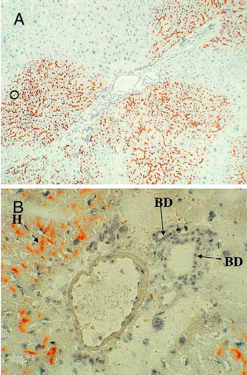
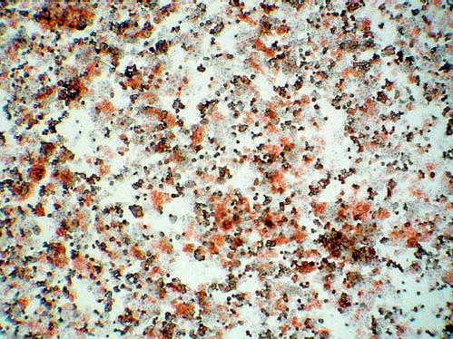
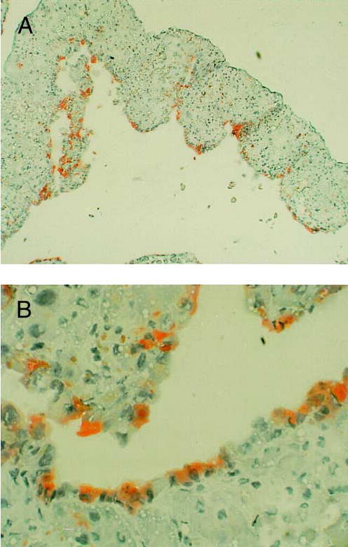
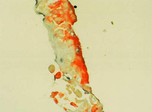
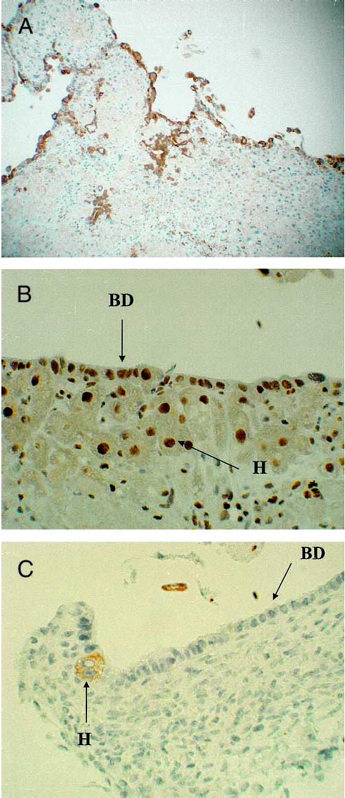
Organoid cultures of hepatocytes in the presence of hepatocyte growth factor (HGF) and epidermal growth factor (EGF) display characteristic histologic organization. Biliary epithelium covers the surface of the tissue exposed to the culture medium. Hepatocytes, stellate cells and endothelial cells compose the underlying structures. In order to investigate the origin of the biliary epithelial cells in the organoid cultures, we utilized the retrorsine/DPPIV system of hepatocyte transplantation to create hybrid livers in which clones of DPPIV hepatocytes colonize variable portions of the lobules. We demonstrate that, as others have shown, biliary epithelium in this in vivo system remains that of the recipient (DPPIV negative) rat. Hepatocytes are the only cells positive for the DPPIV marker enzyme in the hybrid livers. Organoid cultures were prepared from the hybrid livers. Overall, 46.82% of the hepatocytes placed into culture were positive for DPPIV at time zero (after isolation). At 21 days in culture, 47.54% of the biliary epithelium on the surface of the organoid cultures was positive for DPPIV. Since the only DPPIV cells inoculated in the cultures were hepatocytes, this finding demonstrates that, in the conditions of the organoid cultures, hepatocytes do undergo phenotypic transition to biliary epithelial cells.
Figures
Similar articles
-
Contribution of mature hepatocytes to small hepatocyte-like progenitor cells in retrorsine-exposed rats with chimeric livers.Hepatology. 2013 Mar;57(3):1215-24. doi: 10.1002/hep.26104. Epub 2013 Feb 15. Hepatology. 2013. PMID: 23080021
-
Mechanisms of hepatocyte growth factor-mediated and epidermal growth factor-mediated signaling in transdifferentiation of rat hepatocytes to biliary epithelium.Hepatology. 2008 May;47(5):1702-13. doi: 10.1002/hep.22221. Hepatology. 2008. PMID: 18398918 Free PMC article.
-
Histological organization in hepatocyte organoid cultures.Am J Pathol. 2001 Nov;159(5):1877-87. doi: 10.1016/S0002-9440(10)63034-9. Am J Pathol. 2001. PMID: 11696448 Free PMC article.
-
Isolation and culture of biliary epithelial cells.Gut. 1994 Jul;35(7):875-8. doi: 10.1136/gut.35.7.875. Gut. 1994. PMID: 8063212 Free PMC article. Review.
-
Organoid culture to study epithelial cell differentiation and barrier formation in the colon: bridging the gap between monolayer cell culture and human subject research.In Vitro Cell Dev Biol Anim. 2021 Feb;57(2):174-190. doi: 10.1007/s11626-020-00534-6. Epub 2021 Jan 5. In Vitro Cell Dev Biol Anim. 2021. PMID: 33403624 Free PMC article. Review.
Cited by
-
Wnt signaling regulates hepatobiliary repair following cholestatic liver injury in mice.Hepatology. 2016 Nov;64(5):1652-1666. doi: 10.1002/hep.28774. Epub 2016 Sep 26. Hepatology. 2016. PMID: 27533619 Free PMC article.
-
Liver Stem Cells: Experimental Findings and Implications for Human Liver Disease.Gastroenterology. 2015 Oct;149(4):876-882. doi: 10.1053/j.gastro.2015.08.004. Epub 2015 Aug 14. Gastroenterology. 2015. PMID: 26278502 Free PMC article. Review.
-
Hepatocyte growth factor attenuates liver fibrosis induced by bile duct ligation.Am J Pathol. 2006 May;168(5):1500-12. doi: 10.2353/ajpath.2006.050747. Am J Pathol. 2006. PMID: 16651617 Free PMC article.
-
Hepatocyte organoids and cell transplantation: What the future holds.Exp Mol Med. 2021 Oct;53(10):1512-1528. doi: 10.1038/s12276-021-00579-x. Epub 2021 Oct 18. Exp Mol Med. 2021. PMID: 34663941 Free PMC article. Review.
-
Biliary fibrosis drives liver repopulation and phenotype transition of transplanted hepatocytes.J Hepatol. 2016 Jun;64(6):1348-57. doi: 10.1016/j.jhep.2016.01.036. Epub 2016 Feb 5. J Hepatol. 2016. PMID: 26855174 Free PMC article.
References
-
- Evarts RP, Nagy P, Marsden E, Thorgeirsson SS. A precursor-product relationship exists between oval cells and hepatocytes in rat liver. Carcinogenesis. 1987;8:1737–1740. - PubMed
-
- Alison M, Golding M, Lalani EN, Nagy P, Thorgeirsson S, Sarraf C. Wholesale hepatocytic differentiation in the rat from ductular oval cells, the progeny of biliary stem cells. J Hepatol. 1997;26:343–352. - PubMed
-
- Hsia CC, Evarts RP, Nakatsukasa H, Marsden ER, Thorgeirsson SS. Occurrence of oval-type cells in hepatitis B virus-associated human hepato-carcinogenesis. Hepatology. 1992;16:1327–1333. - PubMed
-
- Vandersteenhoven AM, Burchette J, Michalopoulos G. Characterization of ductular hepatocytes in end-stage cirrhosis. Arch Pathol Lab Med. 1990;114:403–406. - PubMed
-
- Haque S, Haruna Y, Saito K, Nalesnik MA, Atillasoy E, Thung SN, Gerber MA. Identification of bipotential progenitor cells in human liver regeneration. Lab Invest. 1996;75:699–705. - PubMed
Publication types
MeSH terms
Substances
Grants and funding
LinkOut - more resources
Full Text Sources
Other Literature Sources