Comparative Study
. 2002 Aug 1;21(15):3927-35.
doi: 10.1093/emboj/cdf410.

Projection structure of the photosynthetic reaction centre-antenna complex of Rhodospirillum rubrum at 8.5 A resolution

Affiliations
Comparative Study

Projection structure of the photosynthetic reaction centre-antenna complex of Rhodospirillum rubrum at 8.5 A resolution

Stuart J Jamieson et al. EMBO J. .

Abstract

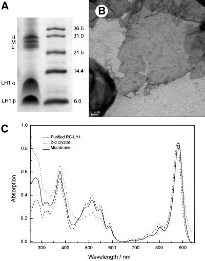
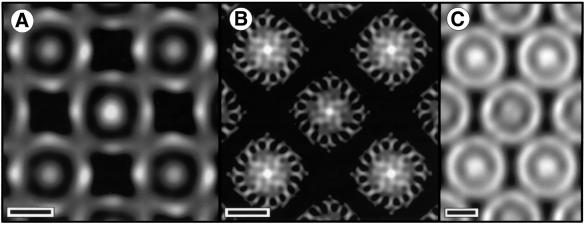
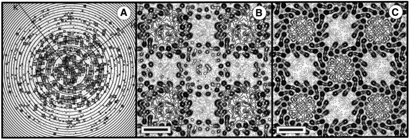
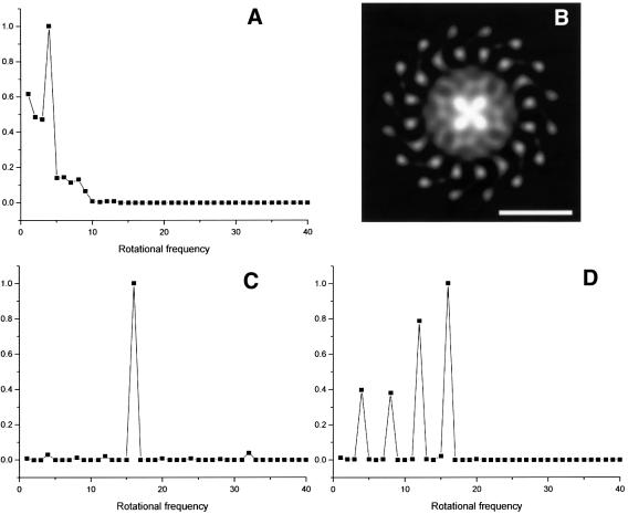
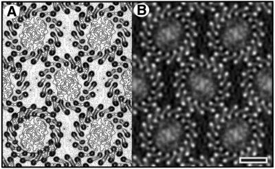
Two-dimensional crystals of the reaction-centre-light-harvesting complex I (RC-LH1) of the purple non- sulfur bacterium Rhodospirillum rubrum have been formed from detergent-solubilized and purified protein complexes. Unstained samples of this intrinsic membrane protein complex have been analysed by electron cryomicroscopy (cryo EM). Projection maps were calculated to 8.5 A from two different crystal forms, and show a single reaction centre surrounded by 16 LH1 subunits in a ring of approximately 115 A diameter. Within each LH1 subunit, densities for the alpha- and beta-polypeptide chains are clearly resolved. In one crystal form the LH1 forms a circular ring, and in the other form the ring is significantly ellipsoidal. In each case, the reaction centre adopts preferred orientations, suggesting specific interactions between the reaction centre and LH1 subunits rather than a continuum of possible orientations with the antenna ring. This experimentally determined structure shows no evidence of any other protein components in the closed LH1 ring. The demonstration of circular or elliptical forms of LH1 indicates that this complex is likely to be flexible in the bacterial membrane.


Figures

None
Fig. 1. (A) Coomassie Blue-stained Schägger gel of 2D crystals of RC–LH1 complex (left column) with subunits labelled. The right column shows the positions of molecular weight standards with molecular weights in kDa. (B) Low-magnification view of a negatively stained 2D crystal. Tetragonal and orthorhombic crystals were indistinguishable at low magnification. (C) Absorbance spectra of chromatophore membranes, purified RC–LH1 complexes and 2D crystals of complexes.
None
Fig. 2. EM in negative stain. Scale bar: 50 Å. (A) Fourier-filtered image of a negatively stained tetragonal crystal. Darker regions correspond to regions of higher density (stain). (B) Tetragonal crystal embedded in a glucose–negative stain mixture. (C) Orthorhombic crystal embedded in negative stain.
None
Fig. 3. (A) Representation of amplitudes of Fourier components calculated for one image of a glucose-embedded tetragonal crystal. Numbers and box sizes correspond to the spot IQ value, with spots of the highest signal-to-noise ratio having an IQ of 1 (Henderson et al., 1986). Spots are shown to a resolution of 1/6 Å–1. (B) 8.5Å resolution projection map calculated from five averaged images of glucose-embedded tetragonal crystals, assuming p1 symmetry. Contouring is at 0.5 r.m.s. density with density above mean (protein) represented by solid contours. Scale bar: 50 Å. (C) As (B), with p4212 symmetry applied.
None
Fig. 4. (A) Rotational power spectrum (Crowther and Amos, 1971) of one of the central RC densities extracted from Figure 3B. Density from the centre of the complex to a radius of 28 Å was analysed. (B) Four-fold rotationally filtered density of one complex extracted from Figure 3B. Whiter regions correspond to higher density. Scale bar: 50 Å. (C) Rotational power spectrum of an entire RC–LH1 complex extracted from Figure 3B. (D) Rotational power spectrum of an outer ring of density of one RC extracted from Figure 3B (radius of 22–39 Å), not including any LH1 density.
None
Fig. 5. (A) 8.5 Å resolution projection map calculated from eight averaged images of glucose-embedded orthorhombic crystals, assuming p22121 symmetry. Exact circles have been drawn through the innermost α- and β-polypeptide densities. Contouring is at 0.5 r.m.s. density with density above mean (protein) represented by solid contours. Scale bar: 50 Å. (B) A grey level representation of the density in (A).
None
Fig. 6. (A) Density for one RC–LH1 complex extracted from Figure 5B. Four prominent density peaks separated by ∼10 Å are marked in red. The 2-fold symmetry-equivalent peaks are not marked. The straight line represents the approximate major axis of the elliptical LH1 ring. (B) Representation of the transmembrane helices of the R.sphaeroides RC aligned to give a best fit with the experimental density. Running from high radius to the centre of the complex, red helices form an arc in the sequence corresponding to LA, LB, LC, LE, LD; likewise, blue helices form an arc in the sequence MA, MB, MC, ME, MD; the yellow helix corresponds to HA; green, ‘special pair’ Bchls; orange, QB. This view is from the cytoplasmic face. The model image was generated with Molscript (Kraulis, 1991) and Raster3D (Merrit and Bacon, 1997). Scale bar: 50 Å.

References

    1. Agarwal R., Rizvi,A.H., Prall,B.S., Olsen,J.D., Hunter,C.N. and Fleming,G.R. (2002) The nature of disorder and inter-complex energy transfer in LH2 at room temperature: a three pulse photon echo peak shift study. J. Phys. Chem B, 18, 624–632.
    1. Allen J.P., Feher,G., Yeates,T.O., Komiya,H. and Rees,D.C. (1987) Structure of the reaction center from Rhodobacter sphaeroides R-26: the cofactors. Proc. Natl Acad. Sci. USA, 84, 5730–5734. - PMC - PubMed
    1. Bellare J.R., Davis,H.T., Scriven,L.E. and Talmon,Y. (1988) Controlled environment vitrification system: an improved sample preparation technique. J. Electron Microsc. Tech., 10, 87–111. - PubMed
    1. Boonstra A.F., Germeroth,L. and Boekema,E. (1994) Structure of the light-harvesting antenna from Rhodospirillum molischianum studied by electron microscopy. Biochim. Biophys. Acta, 1184, 227–234.
    1. Bopp M.A., Sytnik,A., Howard,T.D., Cogdell,R.J. and Hochstrasser,R.M. (1999) The dynamics of structural deformations of immobilized single light-harvesting complexes. Proc. Natl Acad. Sci. USA, 96, 11271–11276. - PMC - PubMed

Publication types

MeSH terms

Substances