. 2002 Aug 1;21(15):3936-48.
doi: 10.1093/emboj/cdf388.

M-LDH serves as a sarcolemmal K(ATP) channel subunit essential for cell protection against ischemia

Affiliations

M-LDH serves as a sarcolemmal K(ATP) channel subunit essential for cell protection against ischemia

Russell M Crawford et al. EMBO J. .

Abstract

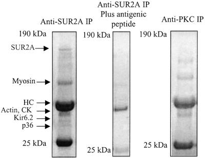
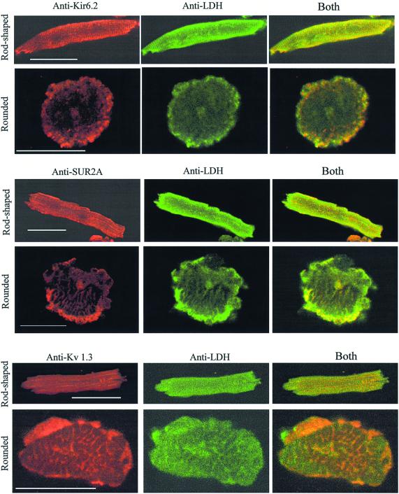
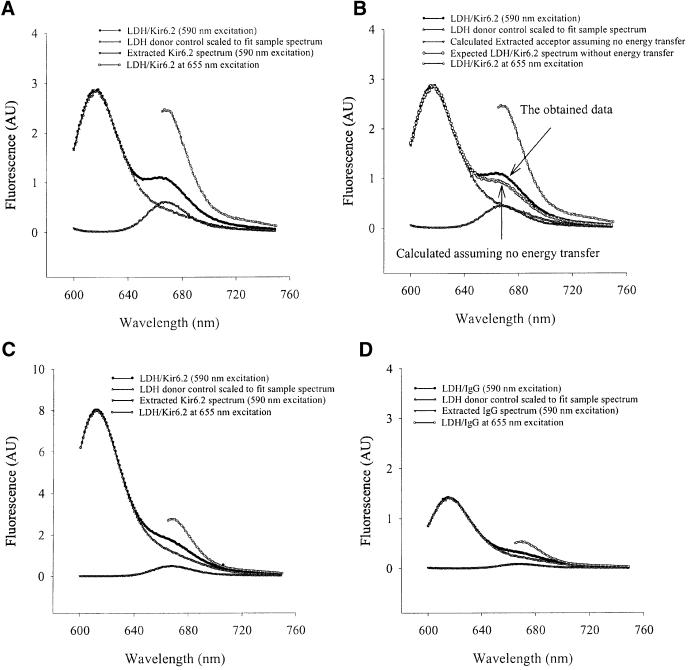
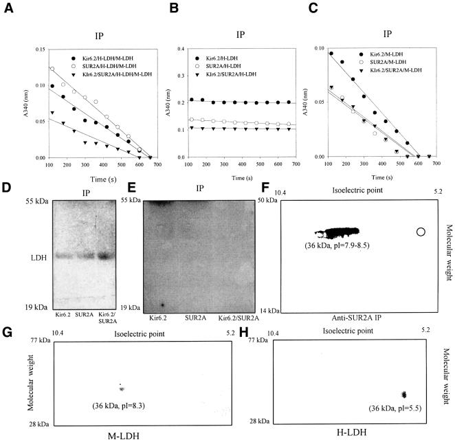
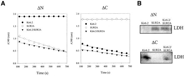
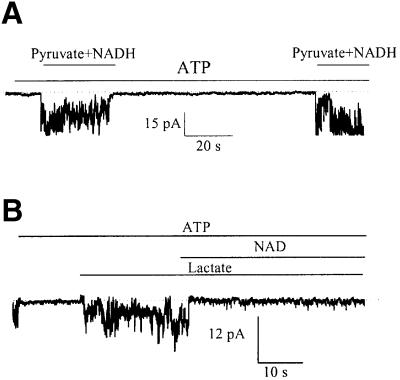
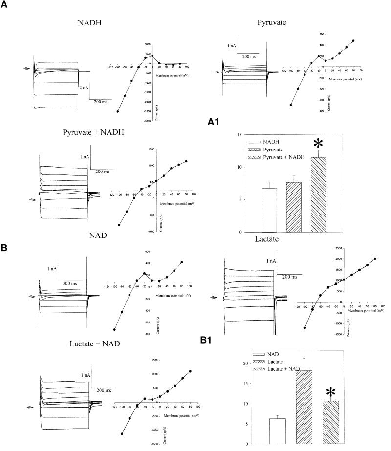
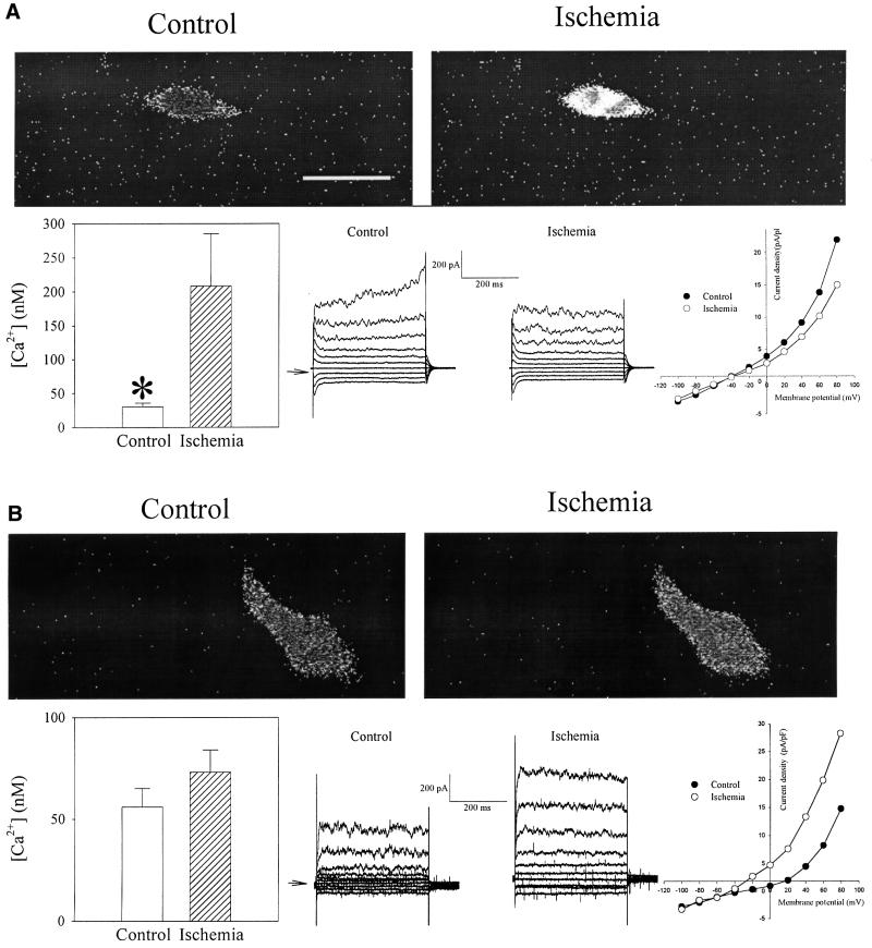
ATP-sensitive K(+) (K(ATP)) channels in the heart are normally closed by high intracellular ATP, but are activated during ischemia to promote cellular survival. These channels are heteromultimers composed of Kir6.2 subunit, an inwardly rectifying K(+) channel core, and SUR2A, a regulatory subunit implicated in ligand-dependent regulation of channel gating. Here, we have shown that the muscle form (M-LDH), but not heart form (H-LDH), of lactate dehydrogenase is directly physically associated with the sarcolemmal K(ATP) channel by interacting with the Kir6.2 subunit via its N-terminus and with the SUR2A subunit via its C-terminus. The species of LDH bound to the channel regulated the channel activity despite millimolar concentration of intracellular ATP. The presence of M-LDH in the channel protein complex was required for opening of K(ATP) channels during ischemia and ischemia-resistant cellular phenotype. We conclude that M-LDH is an integral part of the sarcolemmal K(ATP) channel protein complex in vivo, where, by virtue of its catalytic activity, it couples the metabolic status of the cell with the K(ATP) channels activity that is essential for cell protection against ischemia.


Figures

None
Fig. 1. Immunoprecipitation of cardiac membrane fraction. Coomassie Blue stain of immunoprecipitate pellets (IP) obtained from cardiac membrane fraction precipitated with either anti-SUR2A (without and with antigenic peptide) or anti-PKC antibody.
None
Fig. 2. LDH co-immunoprecipitates with sarcolemmal cardiac KATP channel protein complex and vice versa. (A) MALDI-TOF mass spectrum of tryptic mass fingerprint obtained from an ∼36 kDa migrating protein (identified as LDH). (B) LDH assay with purified LDH (B1) and anti-SUR2A immunoprecipitate of cardiac membrane fraction (B2). (C) Western blotting of anti-Kir6.2 and anti-SUR2A immunoprecipitate of cardiac membrane fraction with anti-LDH antibody. (D) Western blotting of anti-LDH immunoprecipitate (IP) with anti-Kir6.2 and anti-SUR2A antibodies.
None
Fig. 3. Sarcolemmal KATP channel subunits, Kir6.2 and SUR2A, co-localize with LDH in cardiomyocytes. Original images of rod-shaped and rounded cardiomyocytes stained with either anti-Kir6.2 or anti-SUR2A or anti-Kv 1.3 and anti-LDH antibodies labelled with rhodamine (red channel) or fluorescein (green channel) as indicated in the figure. Yellow colour is suggestive of co-localization. Scale bar, 45 µm.
None
Fig. 4. Energy transfer between LDH and Kir6.2. (A) The emission spectra of LDH/Kir6.2, LDH donor control and Kir6.2 acceptor in PBS. (B) The same as in (A) except that the magnitude of the extracted acceptor spectrum is calculated from the (ratio)A value from a sample with only the anti-Kir6.2 antibody. The difference between the sample data and calculated LDH/Kir6.2 spectrum (the sum of donor and acceptor spectra) shows energy transfer. (C) The sample from (A) after the treatment with SDS (1%). (D) The emission spectra of LDH/IgG, LDH donor control and IgG acceptor in PBS. AU, arbitrary units.
None
Fig. 5. The muscle, but not heart, form of LDH directly associates with Kir6.2 and SUR2A subunits. (AC) LDH assay with anti-SUR2A or anti-Kir6.2 immunoprecipitate of untransfected A549 cells or cells transfected with Kir6.2 and SUR2A (alone and in combination) plus M-LDH plus H-LDH (A), H-LDH (B) or M-LDH (C). Anti-Kir6.2 antibody was used for immunoprecipitation of Kir6.2 cells, while for all other groups anti-SUR2A antibody was applied. (D) Western blotting of anti-Kir6.2 and anti-SUR2A immunoprecipitate under conditions in (C). (E) Western blotting of anti-Kir6.2 and anti-SUR2A immunoprecipitate under conditions in (B). (FH) Western blotting 2D gel analysis with anti-LDH antibody of anti-SUR2A immunoprecipitate (IP) of cardiac membrane fraction or purified M- and H-LDH [Sigma; (G) and (H)]. Hollow circle in 2D gel in (F) indicates the expected position for H-LDH signal.
None
Fig. 6. M-LDH associates with Kir6.2 and SUR2A subunits via N- and C-terminus, respectively. (A) LDH assay with anti-SUR2A or anti-Kir6.2 immunoprecipitate of A549 cells transfected with Kir6.2 and SUR2A (alone and in combination) plus N(ΔN)- or C(ΔC)-termini deletion mutants of M-LDH. (B) Western blotting with anti-LDH antibody in conditions under (A).
None
Fig. 7. LDH substrates regulate the opening of KATP channels in excised membrane patches. Recording of KATP channel activity in membrane patch treated with ATP (1 mM) alone, ATP (1 mM) plus pyruvate (20 mM) plus NADH (20 mM), again with ATP (1 mM) alone and again with ATP (1 mM) plus pyruvate (20 mM) plus NADH (20 mM) (A) or treated with ATP (1 mM), ATP (1 mM) plus lactate (20 mM) and ATP (1 mM) plus lactate (20 mM) plus NAD (20 mM) (B). Holding potential –60 mV. Dotted lines correspond to the zero current levels. Both types of experiment were repeated four times with essentially similar results.
None
Fig. 8. LDH substrates regulate KATP channel-mediated membrane current. (A) Membrane currents and corresponding current–voltage relationships in cells filled with pipette solution containing NADH (20 mM) or pyruvate (20 mM) or both. Arrow points to the zero current level. (A1) Current densities at 80 mV for conditions in (A). Vertical bars represent mean ± SEM (n = 6–9). *P <0.01. (B) Membrane currents and corresponding current–voltage relationships in cells filled with pipette solution containing NAD (20 mM) or lactate (20 mM) or both. Arrow points to the zero current level. In all experiments depicted cells were dialysed for ∼5 min. (B1) Current densities at 80 mV under conditions in (B). Vertical bars represent mean ± SEM (n = 6–9). *P <0.05.
None
Fig. 9. Kir6.2/SUR2A/M-LDH confers resistance against ischemia in A549 cells. Original frames and whole-cell recordings under control and ischemic conditions with corresponding bar (intracellular Ca2+ levels, frames) and scatter-line graphs (whole-cell recordings) of (A) untransfected Fura-2 loaded A549 cells and (B) transfected with Kir6.2/SUR2A/M-LDH. Horizontal bar represents 20 µm. Vertical bars represent mean ± SEM, whereas points represent the mean (n = 6–16). *P <0.05.
None
Fig. 10. The opening of KATP channels and intact activity of M-LDH is required for Kir6.2/SUR2A/M-LDH-mediated resistance. Original frames and whole-cell recordings under control and ischemic conditions with corresponding bar (intracellular Ca2+ levels, frames) and scatter-line graphs (whole-cell recordings) of A549 cells transfected with Kir6.2/SUR2A/M-LDH in the presence of 10 µM glybenclamide (A) and transfected with Kir6.2/SUR2A plus 193gly-M-LDH (B) (single-amino acid mutated form of M-LDH with greatly reduced LDH activity; glybenclamide was absent). Scale bar, 20 µm. Vertical bars represent mean ± SEM; points represent mean (n = 5–9). *P <0.05.

References

    1. Altschuld R.A., Jung,D.W., Phillips,R.M., Narayan,P., Castillo,L.C., Whitaker,T.E., Hensley,J., Hohl,C.M. and Brierley,G.P. (1994) Evidence against norepinephrine-stimulated efflux of mitochondrial Mg2+ from intact cardiac myocytes. Am. J. Physiol. Heart Circ. Physiol., 266, H1103–H1111. - PubMed
    1. Ashcroft F.M. and Gribble,F.M. (1998) Correlating structure and function in ATP-sensitive K+ channels. Trends Neurosci., 21, 288–294. - PubMed
    1. Carrasco A.J. et al. (2001) Adenylate kinase phosphotransfer communicates cellular energetic signals to ATP-sensitive potassium channels. Proc. Natl Acad. Sci. USA, 98, 7623–7628. - PMC - PubMed
    1. Clarke A.R., Wigley,D.B., Chia,W.N., Barstow,D., Atkinson,T. and Holbrook,J.J. (1986) Site-directed mutagenesis reveals role of mobile arginine residue in lactate dehydrogenase catalysis. Nature, 324, 699–702. - PubMed
    1. Clegg R.M. (1992) Fluorescence resonance energy transfer and nucleic acids. Methods Enzymol., 211, 353–388. - PubMed

Publication types

MeSH terms