. 2002 Aug 1;21(15):3980-8.
doi: 10.1093/emboj/cdf414.

Intramembrane proteolysis promotes trafficking of hepatitis C virus core protein to lipid droplets

Affiliations

Intramembrane proteolysis promotes trafficking of hepatitis C virus core protein to lipid droplets

John McLauchlan et al. EMBO J. .

Abstract

Hepatitis C virus (HCV) is the major causative pathogen associated with liver cirrhosis and hepatocellular carcinoma. The virus has a positive-sense RNA genome encoding a single polyprotein with the virion components located in the N-terminal portion. During biosynthesis of the polyprotein, an internal signal sequence between the core protein and the envelope protein E1 targets the nascent polypeptide to the endoplasmic reticulum (ER) membrane for translocation of E1 into the ER. Following membrane insertion, the signal sequence is cleaved from E1 by signal peptidase. Here we provide evidence that after cleavage by signal peptidase, the signal peptide is further processed by the intramembrane-cleaving protease SPP that promotes the release of core protein from the ER membrane. Core protein is then free for subsequent trafficking to lipid droplets. This study represents an example of a potential role for intramembrane proteolysis in the maturation of a viral protein.


Figures
None
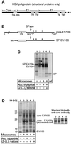
Fig. 1. Analysis of core protein processing in vitro. (A) Diagram of the N-terminal portion of the HCV polyprotein comprising the structural proteins, core, E1 and E2. Sig. seq. indicates the signal sequence at the core-E1 junction; TM, transmembrane regions of the envelope proteins. (B) Diagram of core-E1 constructs used for the in vitro assays. Open bars indicate the hydrophobic region of the signal sequence; arrows, the signal peptidase cleavage site; diamonds, the N-glycosylation sites in E1. (C) Synthesis and processing of SP-E1/100 by in vitro translation in the presence of rough microsomes (lanes 2–4), acceptor tripeptide to inhibit N-glycosylation (lanes 3 and 4), and SPP inhibitor (Z-LL)2- ketone (lane 4). Dots indicate 1x, 2x and 3x glycosylated E1/100; lane 5 shows the reference signal peptide (SP). (D) Synthesis and processing of core-E1/100 by in vitro translation in the presence of rough microsomes (lanes 2–5), acceptor tripepetide to inhibit N-glycosylation (lanes 3 and 5) and SPP inhibitor (lanes 4 and 5). Dots indicate the position of glycosylated E1/100 (E1/100g). The panel to the right shows a corresponding western blot for lanes 2–5 probed with a core-specific antibody.
None
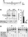
Fig. 2. Mutations affecting processing of the core-E1 signal peptide. (A) Illustration of the mutations in the transmembrane region (underlined) of the signal sequence at the core-E1 junction. Exchanged residues in the mutant, spmt, are indicated. (BIn vitro translation and processing of SP-E1/100 (wt, lanes 1–3) and spmtSP-E1/100 (spmt, lanes 5–7) in the presence of rough microsomes and inhibitor of glycosylation (lanes 2, 3, 6 and 7), and SPP inhibitor (lanes 3 and 7). Lanes 4 and 8 show the respective reference signal peptide (SP). (C) Quanti fication of signal peptide processing using the IQMac v1.2 software (Molecular Dynamics). Grey bars indicate the relative amount of signal peptide obtained in lanes 2 and 6 (no SPP inhibitor) compared with the amount obtained in lanes 3 and 7, respectively, where SPP was inhibited (set to 0% processing). Dark bars indicate the efficiency of processing obtained with the core-E1/100 constructs [(D); lanes 3 wt and spmt]. Relative processing efficiencies were determined accordingly by comparing the amount of processed core protein in lane 3 with that obtained in the presence of SPP inhibitor (lane 4) of the respective panel. Each value was calculated from three independent experiments; the amounts of signal peptide were corrected for variations in translocated E1/100 (for SP-E1/100 constructs). (D) Comparison of core protein produced in cells expressing CE1E2 (wt and spmt) (lane 2) with core protein generated by in vitro translation of core-E1/100 (wt and spmt) in the presence of rough microsomes (lanes 3 and 4) and SPP inhibitor (lane 4). Lanes 1 and 5 show in vitro translated reference peptides corresponding to the N-terminal 179 and 191 residues of the HCV polyprotein, respectively. Dots indicate processed core protein. Note that the wt proteins (left panel) were resolved on Tris–glycine acrylamide gels, which did not resolve spmt proteins of 179 and 191 residues. The latter were analysed on Tris–bicine gels (right panel), where the 191 residue reference peptide showed greater mobility than the shorter 179 residue peptide. After electrophoresis, proteins were transferred to PVDF membranes and probed with a core-specific antibody.
None
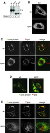
Fig. 3. Trafficking of core protein to lipid droplets requires intra membrane cleavage of the core-E1 signal peptide. CE1E2 polyproteins of the wt and spmt were expressed in BHK cells. (A) Cell extracts were probed for E1 by western blot analysis either directly (lanes 1 and 2) or upon treatment with endoglycosidase H (lanes 3 and 4). (B) Analysis of cells by immunofluorescence with an E2-specific antibody. (C) Immunofluorescence of core protein combined with Oil Red O staining of lipid droplets. (D) As for (C) except that a wider field is shown. (E) As for (C) except that the cells used were Huh7 cells.
None
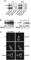
Fig. 4. Mutations affecting intramembrane cleavage of the signal peptide at the core-E1 junction retain core protein at the ER membrane. (A) Wt and spmt CE1E2 polyproteins were expressed in BHK cells. Membranes were isolated from cells and extracted with sodium carbonate (lanes 3 and 4). The distribution of core protein and E2 were visualized by western blot analysis with a core- and an E2-specific antibody, respectively. (B) CE1E2 (lane 1), ΔCE1E2 (lanes 2 and 3) and ΔCspmtE1E2 (lanes 4 and 5) polyproteins were expressed in BHK cells in the absence or presence of proteasome inhibitor MG132 as indicated. Membranes were isolated and extracted with sodium carbonate, and the distributions of E2 (left panel) and core/ΔC protein (right panel) were visualized by western blot analysis with a core- and an E2-specific antibody, respectively. (C) Immunofluorescence of cells probed with a core- and an E2-specific antibody. Where indicated, proteasome inhibitor MG132 was present during expression of ΔCE1E2.
None
Fig. 5. Model for the release of HCV core protein from the ER membrane and its association with lipid droplets. (A) The central hydrophobic domain 2 (triangle) of HCV core protein anchors the protein in the phospholipid monolayer surrounding lipid droplets. During biosynthesis of the HCV polyprotein, core protein is cleaved from the nascent polypeptide by signal peptidase. Subsequent processing by SPP within the centre of the membrane-spanning portion of the signal peptide (barrel) liberates core protein from the membrane. Core protein, with domain 2 integrated into the cytosolic leaflet of the bilayer, is now free for entering the zone of lipid droplet formation. TG, triacylglycerol. (B) Mutant ΔC, which lacks domain 2, is targeted to the ER membrane and cleaved by signal peptidase as wt core protein. Processing by SPP liberates ΔC from the ER membrane leading to degradation by the proteasome.

Similar articles

Cited by

References

    1. Amberg S.M. and Rice,C.M. (1999) Mutagenesis of the NS2B-NS3-mediated cleavage site in the flavivirus capsid protein demonstrates a requirement for coordinated processing. J. Virol., 73, 8083–8094. - PMC - PubMed
    1. Barba G. et al. (1997) Hepatitis C virus core protein shows a cytoplasmic localization and associates to cellular lipid storage droplets. Proc. Natl Acad. Sci. USA, 94, 1200–1205. - PMC - PubMed
    1. Beard M.R. et al. (1999) An infectious molecular clone of a Japanese genotype 1b hepatitis C virus. Hepatology, 30, 316–324. - PubMed
    1. Brown D.A. (2001) Lipid droplets: proteins floating on a pool of fat. Curr. Biol., 11, R446–R449. - PubMed
    1. Bukh J., Purcell,R.H. and Miller,R.H. (1994) Sequence analysis of the core gene of 14 hepatitis C virus genotypes. Proc. Natl Acad. Sci. USA, 91, 8239–8243. - PMC - PubMed

Publication types

MeSH terms

Substances