. 2002 Aug 1;21(15):4001-11.
doi: 10.1093/emboj/cdf393.

A large dispersed chromosomal region required for chromosome segregation in sporulating cells of Bacillus subtilis

Affiliations

A large dispersed chromosomal region required for chromosome segregation in sporulating cells of Bacillus subtilis

Ling Juan Wu et al. EMBO J. .

Abstract

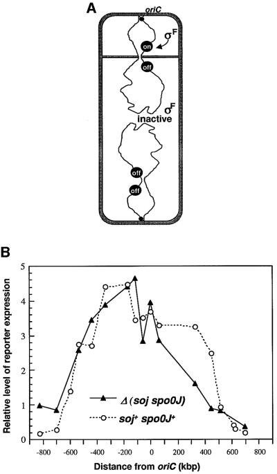
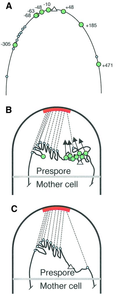
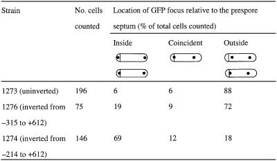
The cis-acting sequences required for chromosome segregation are poorly understood in most organisms, including bacteria. Sporulating cells of Bacillus subtilis undergo an unusual asymmetric cell division during which the origin of DNA replication (oriC) region of the chromosome migrates to an extreme polar position. We have now characterized the sequences required for this migration. We show that the previously characterized soj-spo0J chromosome segregation system is not essential for chromosome movement to the cell pole, so this must be driven by an additional segregation mechanism. Observations on a large set of precisely engineered chromosomal inversions and translocations have identified a polar localization region (PLR), which lies approximately 150-300 kbp to the left of oriC. Surprisingly, oriC itself has no involvement in this chromosome segregation system. Dissection of the PLR showed that it has internal functional redundancy, reminiscent of the large diffuse centromeres of most eukaryotic cells.


Figures

None
Fig. 1. Effect of deletion of the soj–spo0J locus on chromosome trapping in the prespore compartment. (A) Schematic representation of the reporter trapping system. Soon after the onset of sporulation an asymmetrically positioned division septum closes around one of the two chromosomes of the cell to form the prespore compartment. SpoIIIE protein then normally drives transfer of the remainder of the chromosome into the small compartment but in spoIIIE mutants the chromosome remains in this bisected state. The prespore chromosome has a relatively fixed orientation with the oriC region invariably located within the prespore compartment and oriC-distal sequences located outside in the mother cell. A prespore-specific transcription factor, σF, becomes active in the small compartment and can drive expression of a σF-dependent reporter gene if located in the segment of the chromosome that is trapped in the prespore (‘on’) but not if it is located in the oriC-distal part of the chromosome (‘off’). (B) Effect of soj–spo0J deletion on the accumulation of β-galactosidase expressed from the σF-dependent gpr–lacZ reporter at different chromosomal positions (in a spoIIIE36 background). Strains were induced to sporulate and assayed at intervals for β-galactosidase activity. Open circles show results obtained previously with soj+ spo0J+ cells (Wu and Errington, 1998). Filled triangles show the data for the soj–spo0J(yyaA–yyaC)::kan] deletion derivatives (average of two to eight experiments, in samples taken after 3 h of sporulation, representative of the full time courses). The values given have been normalized to the activities obtained with the equivalent spoIIIE+ strains (set at 1), induced to sporulate and sampled in parallel (see Wu and Errington, 1998).
None
Fig. 2. Effect of defined chromosome inversions on the segment of the chromosome localized in the prespore. (A) Schematic of the method used to construct defined chromosome inversions. The left and right arms of the circular chromosome are represented in hatched and solid lines, respectively. These are bounded at oriC (the origin of bi-directional chromosome replication) and terC (the terminus of replication). Various inserted resistance genes are indicated as follows: spc, spectinomycin; tet, tetracycline; neo, neomycin. ‘neo, neo’ and ‘neo’ represent non-functional derivatives of the neo gene truncated at the 5′, 3′, or 5′ and 3′ ends, respectively. Homologous recombination between the truncated neo genes in the left and right chromosome arms results in a chromosome inversion in which the upper part of the chromosome, containing oriC, is switched around relative to the lower part. Note that this does not change the direction of DNA replication through the chromosome arms. (B) Locations of the inversion end points (numbered) and the σF-dependent gpr–lacZ reporters (blue ovals). (C) β-galactosidase accumulation in different inversion strains. Two independently isolated strains with inversions from ycgA (+325) to various locations on the left arm of the chromosome (‘Inverted’) were patched onto agar plates containing X-Gal, together with the un-inverted strains (‘Un-inverted’) containing the inversion elements and the parent strain (‘Parent’) 1265 [spoIIIE36 Δ(yyaA–yyaC)::erm amyE::gpr–lacZ] or 1266 [spoIIIE36 Δ(yyaA–yyaC)::erm spoIID::gpr–lacZ], and grown at 37°C for 3 days. The left panel shows the strains with the σF-dependent gpr–lacZ fusion at the spoIID locus, and the right panel shows strains with the fusion at amyE. The locations of the inversion end points on the left chromosome arm are shown in the middle. (D) β-galactosidase accumulation in a subset of inversion strains bearing a spoIIQ–lacZ fusion at the amyE locus. Inversion end points labelled as for (C). (EG) Quantitative analysis of reporter expression during sporulation in liquid culture. The strains containing gpr–lacZ at amyE [(E), before inversion; (F), after inversion] or at spoIID [(G), after inversion] were induced to sporulate at time zero, and samples were taken at intervals for assay of β-galactosidase activity. A single experiment representative of several repeats is shown.
None
Fig. 3. Summary of the effect of inversions at a distant location (+612) on chromosome trapping as measured by the accumulation of β-galactosidase from the σF-dependent gpr–lacZ reporter located at six different chromosomal positions. Strains were patched to and grown on agar plates containing X-Gal as described in the legend to Figure 2. The activities of the reporter in a series of experiments were scored and depicted as circles with different degrees of shading at the corresponding locations. The top line (labelled wt) shows the oriC region of the wild-type chromosome. The right arm of the chromosome is depicted with a thin black line. The left arm is depicted with a thick line, with different segments, demarcated by the different inversion sites used, shown in different colours. The positions of the different gpr–lacZ reporter insertions are shown as circles, with the corresponding level of β-galactosidase activity indicated by the shading (scored on a scale of 1 to 5 as indicated). Lines labelled (i)–(vi) below show the chromosomal structures of the different inversion strains tested, with the inversion end points indicated by black arrowheads. The location of the putative PLR is indicted by the boxed region (though some polar localizing activity clearly lies outside this region).
None
Fig. 4. Fluorescence microscopy showing sporulating cells with a lacO array at dinB (+608) before [(A), strain 1273] and after [(B), strain 1274] chromosome inversion from –214 to +612 kbp. The strains were induced to sporulate, stained with the membrane dye FM5-95 and viewed under fluorescence microscope as described in Materials and methods. Left panels show the cell outline and the asymmetric septa as revealed by the membrane dye. Right panels are GFP signals showing the cellular location of the lacO. It also shows faint staining of the membrane because the signals from the dye crossed into the GFP channel. Arrows point to the positions of asymmetric septa.
None
Fig. 5. A model for prespore chromosome segregation and possible roles for the Soj–Spo0J system. (A) Layout of known Spo0J-binding sites (labelled large green circles; as defined by Lin and Grossman, 1998) and putative PLR sites (small blue circles) in the oriC (white triangle) region of the chromosome. The segment of DNA depicted (thin black line) shows approximately the segment of DNA that is normally trapped in the prespore compartment of spoIIIE mutant cells (soj+ spo0J+) during sporulation (approximately +600 to –700; see Figure 1B). On the basis of our results, the hypothetical PLR sites mainly lie in the –149 to –315 region. (B) Association of proteins bound to sites in the PLR with a recognition site or anchor (red bar) in the vicinity of the cell pole during sporulation results in dragging of the PLR towards the cell pole (dotted black lines). oriC itself is efficiently trapped in the prespore because Spo0J protein forms a complex that condenses the oriC region in close juxtaposition to the PLR. Alternatively or additionally, Spo0J may be directly attracted towards the cell pole, as indicated by the arrows. (C) Effect of disruption of the soj–spo0J system on trapping of DNA in the prespore compartment.
None

References

    1. Anagnostopoulos C. and Spizizen,J. (1961) Requirements for transformation in Bacillus subtilis. J. Bacteriol., 81, 741–746. - PMC - PubMed
    1. Autret S., Nair,R. and Errington,J. (2001) Genetic analysis of the chromosome segregation protein Spo0J of Bacillus subtilis: evidence for separate domains involved in DNA binding and interactions with Soj protein. Mol. Microbiol., 41, 743–755. - PubMed
    1. Bath J., Wu,L.J., Errington,J. and Wang,J.C. (2000) Role of Bacillus subtilis SpoIIIE in DNA transport across the mother cell–prespore division septum. Science, 290, 995–997. - PubMed
    1. Bylund J.E., Haines,M.A., Piggot,P.J. and Higgins,M.L. (1993) Axial filament formation in Bacillus subtilis: induction of nucleoids of increasing length after addition of chloramphenicol to exponential-phase cultures approaching stationary phase. J. Bacteriol., 175, 1886–1890. - PMC - PubMed
    1. Cervin M.A., Spiegelman,G.B., Raether,B., Ohlsen,K., Perego,M. and Hoch,J.A. (1998) A negative regulator linking chromosome segregation to developmental transcription in Bacillus subtilis. Mol. Microbiol., 29, 85–95. - PubMed

Publication types

MeSH terms