Comparative Study
. 2002 Aug 1;21(15):4026-36.
doi: 10.1093/emboj/cdf400.

Mos is not required for the initiation of meiotic maturation in Xenopus oocytes

Affiliations
Comparative Study

Mos is not required for the initiation of meiotic maturation in Xenopus oocytes

Aude Dupré et al. EMBO J. .

Abstract

In Xenopus oocytes, the c-mos proto-oncogene product has been proposed to act downstream of progesterone to control the entry into meiosis I, the transition from meiosis I to meiosis II, which is characterized by the absence of S phase, and the metaphase II arrest seen prior to fertilization. Here, we report that inhibition of Mos synthesis by morpholino antisense oligonucleotides does not prevent the progesterone-induced initiation of Xenopus oocyte meiotic maturation, as previously thought. Mos-depleted oocytes complete meiosis I but fail to arrest at metaphase II, entering a series of embryonic-like cell cycles accompanied by oscillations of Cdc2 activity and DNA replication. We propose that the unique and conserved role of Mos is to prevent mitotic cell cycles of the female gamete until the fertilization in Xenopus, starfish and mouse oocytes.


Figures
None
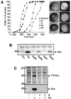
Fig. 1. Morpholino antisense inhibits Mos synthesis but does not prevent GVBD. (A) Prophase oocytes were injected or not (solid squares) with either 130 ng of antisense oligonucleotides A (open circles), 130 ng of control oligonucleotides Ac (solid circles), 84 ng of morpholino antisense M (open triangles) or 84 ng of control morpholino Mc (solid triangles). One hour after injection, oocytes were induced to mature by progesterone (Pg) and the percentage of GVBD was determined as a function of time by following the appearance of the typical white spot at the animal pole of the oocyte (left panel). Right panel: first row illustrates the external morphology of oocytes and second row illustrates sections of fixed oocytes (Pro, prophase oocyte; Pg, progesterone-matured oocyte at GVBD; Pg/M, morpholino antisense-injected oocyte treated by progesterone and fixed at GVBD). Arrowhead indicates the GV. (B) Western blots using Xenopus Mos antibody. One oocyte from the previous experiment was homogenized at GVBD and loaded in each lane. One prophase oocyte (Pro) was loaded on the first lane. The position of one molecular weight marker is indicated on the left. (C) Groups of 50 prophase oocytes were microinjected or not with morpholino antisense (M) and were metabolically labeled with [35S]methionine at 200 µCi/ml. One hour later, progesterone (Pg) was added or not. Progesterone-treated oocytes were selected 4 h after GVBD. Mos immunoprecipitates were electrophoresed, transferred to nitrocellulose and first exposed to autoradiography (upper panel) and then probed with anti-Mos antibody (lower panel). The positions of molecular weight markers are indicated on the left.
None
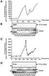
Fig. 2. Progesterone-induced Cdc2 activation in Mos-ablated oocytes. Oocytes were injected (C and D) or not (A and B) with morpholino antisense. One hour later (time 0), progesterone was added and oocytes were collected and homogenized at indicated times. eGVBD, first pigment rearrangement detected at the animal pole (15 min before GVBD); GVBD, well-defined white spot observed at 225 min in control oocytes and at 360 min in morpholino antisense-injected oocytes. The equivalent of three oocytes was assayed for H1 kinase activity (A and C). At indicated times, one oocyte was immunoblotted with antibodies against Tyr15-phosphorylated Cdc2 or against Cdc25 phosphatase (B and D). The positions of the molecular weight markers are indicated on the left.
None
Fig. 3. Cyclin B degradation is not affected in the absence of Mos. Oocytes were injected or not with morpholino antisense (M). One hour later (time 0), progesterone (Pg) was added and oocytes were homogenized at the indicated times. One oocyte equivalent was loaded in each lane and immunoblots were performed with antibodies against (AXenopus Cyclin B2, (BXenopus Cyclin B1 and (C) Cdc27. Oocyte lysates were originated from the experiment already described in Figure 2. The positions of the molecular weight markers are indicated on the left.
None
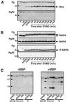
Fig. 4. Mos ablation prevents MAPK activation induced by progesterone. Oocytes were injected or not with morpholino antisense (M) and incubated in the presence of progesterone (Pg) 1 h later (time 0). Oocytes were collected at indicated times and lysates originated from the experiment illustrated in Figure 2 were immunoblotted with the antibodies directed against (AXenopus Mos and (B) total MAPK or the active phosphorylated form of MAPK (P-MAPK). The positions of the molecular weight markers are indicated on the left. (C) In-gel assay of MBP kinase activities. Oocytes were injected or not with morpholino antisense (M) or incubated in the presence of 50 µM U0126. One hour after injection or 20 min after incubation in U0126, progesterone was added (time 0) and oocytes were collected either at time 0 or at GVBD or 4 h after GVBD (240). MBP kinase activity was measured by an in-gel assay after adding (+MBP) or not (–MBP) myelin basic protein in the gel. The equivalent of one oocyte was loaded per lane. The positions of the molecular weight markers are indicated on the left.
None
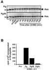
Fig. 5. Rsk is partially activated in the absence of Mos and MAPK activation. (A) Oocytes were injected or not with morpholino antisense (M). One hour later, progesterone (Pg) was added (time 0) and oocytes were homogenized at the indicated times. Oocyte extracts originated from the experiment illustrated in Figure 2 were immunoblotted with an antibody against Rsk2. (B) Oocytes were injected or not with either morpholino antisense (M) or traditional antisense (A) oligonucleotides and were incubated in the presence of progesterone (Pg). Groups of 10 oocytes, either at prophase stage (Pro) or 4 h after GVBD (GVBD + 4 h) were homogenized and immunoprecipitated with the anti-Rsk2 antibody. Kinase activity was assayed using S6 peptide in immunoprecipitates.
None
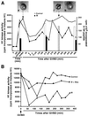
Fig. 6. Mos is required to stabilize Cdc2 activity after GVBD. (A) Oocytes were injected (solid squares, solid bars) or not (open squares, open bars) with morpholino antisense (M). One hour later (time 0), progesterone was added. GVBD started 4 h after progesterone addition in control oocytes, and with a 4 h delay in morpholino antisense-injected oocytes. At GVBD, some morpholino antisense-injected oocytes were injected with 50 ng of recombinant MBP–Mos protein (hatched bar). At indicated times, oocytes were homogenized and kinase activities of Cdc2 (H1 phosphorylation: lines) and Rsk (S6 peptide phosphorylation: bars) were assayed. Each point of Cdc2 and Rsk activities corresponds to four oocytes. The external morphology of injected oocytes is illustrated at the time of GVBD (a) and 5.5 h after GVBD with (b) or without (c) MBP–Mos injection at GVBD. (B) Oocytes were injected (solid squares) or not (open squares) with morpholino antisense (M). One hour later (time 0), progesterone was added. GVBD started 4 h after progesterone addition in control oocytes, and with 3 h delay in morpholino antisense-injected oocytes. At GVBD, some Mos-ablated oocytes were injected with 50 ng of recombinant MBP–Mos protein (triangles). At indicated times, oocytes were homogenized and Cdc2 kinase activity was assayed (three oocytes per point).
None
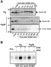
Fig. 7. Analysis of Cyclin B2 levels, Cdc2 phosphorylation and DNA replication in Mos-ablated oocytes after GVBD. (A) Oocytes were injected or not with morpholino antisense (M). Six hours later, progesterone (Pg) was added (time 0). Some of the progesterone-treated oocytes were incubated 45 min after GVBD in CHX. Oocytes were collected at indicated times and immunoblotted with antibodies against Xenopus Cyclin B2 and Tyr15-phosphorylated form of Cdc2. GVBD occurred 5 h after progesterone addition in control oocytes and 3 h later in morpholino antisense-injected oocytes. (B) In parallel, 1 h after progesterone addition, oocytes were injected with [α-32P]dCTP (50 nl, 3000 Ci/mmol). Aphidicolin (APD) was added at the time of GVBD and CHX 45 min after GVBD. Five hours after GVBD, DNA was extracted and incorporation of radioactive dCTP was visualized by autoradiography. Each lane corresponds to the equivalent of one oocyte. Prophase oocytes (Pro) were also injected with [α-32P]dCTP and collected 10 h after GVBD occurring in the control oocytes.

Similar articles

Cited by

References

    1. Ballantyne S., Daniel,D.L. and Wickens,M. (1997) A dependent pathway of cytoplasmic polyadenylation reactions linked to cell cycle control by c-mos and CDK1 activation. Mol. Biol. Cell, 8, 1633–1648. - PMC - PubMed
    1. Bhatt R.R. and Ferrell,J.E.,Jr (1999) The protein kinase p90rsk as an essential mediator of cytostatic factor activity. Science, 286, 1362–1365. - PubMed
    1. Choi T., Rulong,S., Resau,J., Fukasawa,K., Matten,W., Kuriyama,R., Mansour,S., Ahn,N. and Van de Woude,G.F. (1996) Mos/mitogen-activated protein kinase can induce early meiotic phenotypes in the absence of maturation-promoting factor—a novel system for analyzing spindle formation during meiosis I. Proc. Natl Acad. Sci. USA, 93, 4730–4735. - PMC - PubMed
    1. Colledge W.H., Carlton,M.B.L., Udy,G.B. and Evans,M.J. (1994) Disruption of c-mos causes parthenogenetic development of unfertilized mouse eggs. Nature, 370, 65–68. - PubMed
    1. Daar I., Paules,R.S. and Van de Woude,G.F. (1991) A characterization of cytostatic factor activity from Xenopus eggs and c-mos-transformed cells. J. Cell Biol., 114, 329–335. - PMC - PubMed

Publication types

MeSH terms