Comparative Study
. 2002 Aug 1;21(15):4125-35.
doi: 10.1093/emboj/cdf390.

Yhh1p/Cft1p directly links poly(A) site recognition and RNA polymerase II transcription termination

Affiliations
Comparative Study

Yhh1p/Cft1p directly links poly(A) site recognition and RNA polymerase II transcription termination

Bernhard Dichtl et al. EMBO J. .

Abstract

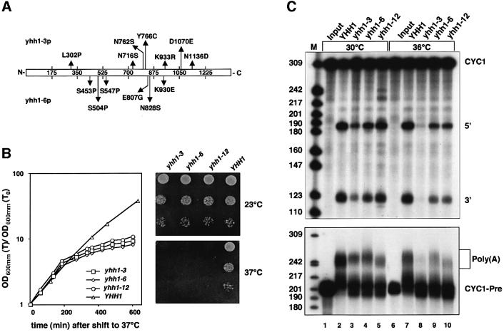
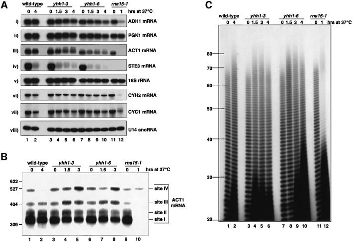
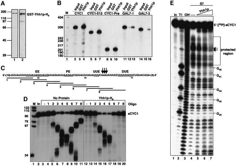
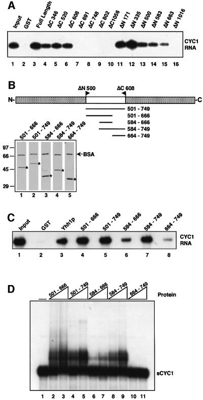
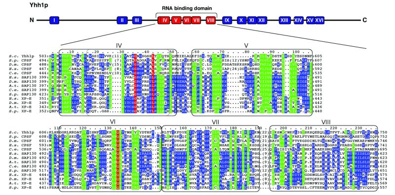
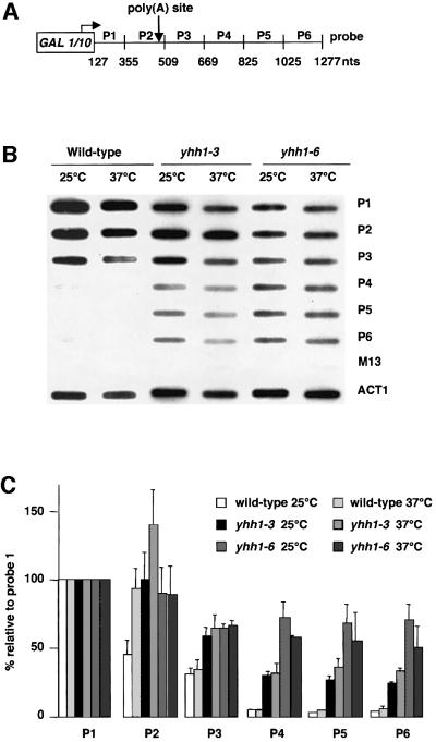
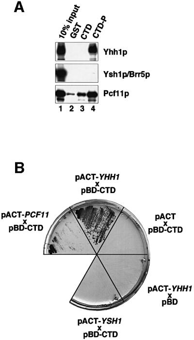
RNA polymerase II (pol II) transcription termination requires co-transcriptional recognition of a functional polyadenylation signal, but the molecular mechanisms that transduce this signal to pol II remain unclear. We show that Yhh1p/Cft1p, the yeast homologue of the mammalian AAUAAA interacting protein CPSF 160, is an RNA-binding protein and provide evidence that it participates in poly(A) site recognition. Interestingly, RNA binding is mediated by a central domain composed of predicted beta-propeller-forming repeats, which occurs in proteins of diverse cellular functions. We also found that Yhh1p/Cft1p bound specifically to the phosphorylated C-terminal domain (CTD) of pol II in vitro and in a two-hybrid test in vivo. Furthermore, transcriptional run-on analysis demonstrated that yhh1 mutants were defective in transcription termination, suggesting that Yhh1p/Cft1p functions in the coupling of transcription and 3'-end formation. We propose that direct interactions of Yhh1p/Cft1p with both the RNA transcript and the CTD are required to communicate poly(A) site recognition to elongating pol II to initiate transcription termination.


Figures

None
Fig. 1. Analysis of pre-mRNA 3′-end processing activity of yhh1 mutant extracts in vitro. (A) Sequence analysis of yhh1 mutant alleles revealed seven amino acid changes in yhh1-3p (top of the panel) and six amino acid changes in yhh1-6p (bottom of panel), which underlie the respective ts phenotypes. (B) Growth curves of wild-type and mutant YHH1 strains after shift to 37°C. Ten-fold serial dilutions of cultures spotted on YPD plates followed by incubation at the indicated temperatures for 2 days. (CIn vitro cleavage (upper panel) and polyadenylation (lower panel) assays with extracts prepared from yeast strains as indicated. Input lanes represent mock-treated reactions. Length of marker bands is indicated on the left. For 3′-end cleavage, internally 32P-labelled CYC1 RNA was used. The positions of full-length RNA (CYC1), 5′ and 3′ cleavage products are indicated. Cleavage was performed either at 30°C (lanes 1–5) or at 36°C (lanes 6–10). Specific polyadenylation was performed with internally 32P-labelled CYC1-precleaved RNA that ends at the natural cleavage site. The positions of substrate (CYC1-Pre) and polyadenylation products [Poly(A)] are indicated. Polyadenylation assays were performed either at 30°C (lanes 1–5) or at 37°C, following a 5 min pre-incubation of extract and reaction mixture at this temperature (lanes 6–10).
None
Fig. 2. yhh1 mutant cells are defective in recognition of the ACT1 poly(A) site. (A) Northern analysis of total RNA extracted from wild-type and mutant YHH1 cells grown at 23°C, or following a shift to 37°C for 1.5, 3 and 4 h. For control, an rna15-1 strain grown at 23°C and 37°C was analysed in parallel. RNAs were separated on formaldehyde/1.2% agarose gels (panels i–v) or 8.3 M urea/8% polyacrylamide gels (panels vi–viii). Filters were developed with random-primed labelled probes or end-labelled oligonucleotides directed against the RNA species indicated on the right of each panel. (B) Analysis of ACT1 poly(A) site usage in wild-type and yhh1 and rna15-1 mutant cells as indicated. Total RNAs extracted from strains grown as described in (A) were treated with RNase H and oligonucleotide ACT1-RNaseH, and analysed by northern blotting. A random-primed probe complementary to the ACT1 3′-end region (nucleotides 930–1536) was used. The positions of RNAs polyadenylated at sites I to IV are indicated, as well as molecular weight markers. (C) Poly(A) tail analysis with total RNA extracted from indicated YHH1 strains after growth at 23°C (0 h) or after shift to 37°C for 1.5, 3 and 4 h. Also analysed was an rna15-1 strain grown at 23°C or after shift to 37°C for 1 h. Poly(A) tail length is indicated on the left. Strong signals of short RNA species in lanes 2, 4, 5, 6, 9, 10 and 12 were not reproducible and probably result from incomplete digestion of non-poly(A) sequences.
None
Fig. 3. Yhh1p is an RNA-binding protein. (A) Purified GST-Yhh1p-H6 expressed in E.coli was subjected to SDS–PAGE and visualized by Coomassie Brilliant Blue staining (lane 1) or detected after western transfer with an anti-Yhh1p serum (lane 2). Positions of recombinant protein and marker bands are indicated. (B) GST pull-down experiments with 0.5 µg GST (lanes 3, 6, 9, 12 and 15) or GST-Yhh1p-H6 (Yhh1p; lanes 4, 7, 10, 13 and 16) and in vitro transcribed, 32P-labelled RNAs. Lanes 2, 5, 8, 11 and 14 show 10% of RNA included in binding reactions (Input). Length of marker bands (M; lane 1) is indicated on the left. (C) Sequence of sCYC1 RNA. Only CYC1-derived nucleotides are shown; vector encoded nucleotides are indicated by numbers in parentheses. Efficiency (EE) and positioning elements (PE), and upstream (UUE) and downstream (DUE) U-rich elements are underlined. The arrows mark poly(A)-site positions. Sequences complementary to 14-mer DNA oligonucleotides (1–8) are indicated. (D) RNase H protection analysis with Yhh1p and sCYC1 RNA. Internally 32P-labelled sCYC1 RNA was incubated with binding buffer (No protein; lanes 2–11) or GST-Yhh1p-H6. The GST moiety was removed from recombinant protein by cleavage with TEV protease included in the binding reaction. Protein containing assays are therefore indicated by Yhh1p-H6 (lanes 12–20). After pre-incubation, the substrate was cleaved by addition of RNase H and oligonucleotides as indicated. For control, oligonucleotides were omitted in lanes 3 and 12. The RNA input (In) is shown in lane 2. HpaII-digested pBR322 fragments were 5′-end labelled and served as marker bands (M; lane 1). (E) S7 nuclease protection analysis. 5′-[32P]-labelled sCYC1 RNA was pre-incubated in the absence (lane 4) and presence of increasing amounts of GST-Yhh1p-His6 (100, 200 and 400 ng; lanes 5–7), followed by the addition of S7 nuclease. Reaction products were separated on 12% polyacrylamide/8.3 M urea gels. Also analysed were the RNA input (In; lane 1), a partial T1 nuclease digest (T1; T1 cuts RNA after guanosines) and a partial alkaline hydrolysis of the input RNA (OH; lane 3). To the right of the panel, the migration of full-length RNA and the position of guanosines in the RNA substrate are indicated. Arrows mark the in vitro cleavage sites and the region protected by GST-Yhh1p-His6 is indicated.
None
Fig. 4. Delineation of the Yhh1p RNA-binding domain. (A) GST pull-down with in vitro transcribed, 32P-labelled CYC1 RNA and 0.5 µg GST (lane 2), GST-Yhh1p-H6 (Yhh1p; lane 3), C-terminal (lanes 4–10) and N-terminal truncations of Yhh1p (lanes 11–16) as indicated on top of the panel. Lane 1 shows 10% of RNA in binding reactions (Input). (B) Representation of internal Yhh1p domains that were expressed as N-terminal GST fusions and with a C-terminal His6 tag. For quality control, recombinant proteins (indicated by an asterisk) were resolved by SDS–PAGE and stained with Coomassie Brilliant Blue. Migration of BSA that was included in the final elution buffer is indicated. (C) 32P-labelled CYC1 RNA was tested for interaction with 0.5 µg GST (lane 2), GST-Yhh1p-H6 (Yhh1p; lane 3), and Yhh1p domains as indicated in (B) (lanes 4–8). Lane 1 shows 10% of RNA in binding reactions (Input). (D) Mobility shift experiments with Yhh1p domains as indicated in (B). 32P-labelled sCYC1 RNA was incubated with increasing amounts (200 and 400 ng) of protein and analysed by native polyacrylamide gel electrophoresis. Protein was omitted in lane 1. The position of sCYC1 is indicated.
None
Fig. 5. Alignment of sequences related to the Yhh1p RNA-binding domain. Sequences similar to the RNA-binding domain of Yhh1p were aligned as described in Materials and methods. Invariant residues are shown in red; green indicates conserved and similar amino acid positions which are present in 70–100% of the aligned sequences, and conserved and similar residues occurring in 35–70% of the aligned sequences are shown in blue. Also indicated are sequence repeats within Yhh1p (boxes numbered I–XVI) that were predicted to form β-propeller-like structures (Neuwald and Poleksic, 2000). The sequence repeats that align with the active RNA-binding fragments of Yhh1p (aa 501–666, repeats IV–VII, and aa 584–749, repeats V–VIII) are shown in red. DDBJ/EMBL/Genbank accession numbers are: Homo Sapiens (H.s.) SAP130: CAB56791; Drosophila melanogaster (D.m.) SAP130: AAF47416; Arabidopsis thaliana (A.t.) SAP130: CAB75754; Caenorhabditis elegans (C.e.) SAP130: T32916; S.pombe (S.p.) SAP130: BAA86918; H.s. XP-E: CAA05770; A.t. XP-E: CAB81084; D.m. XP-E: AAF54901; S.p. XP-E: T37876; D.m. CPSF: AAF58240; H.s. CPSF: Q10570; C.e. CPSF: AAF36067; S.p. CPSF: T39636; S.cerevisiae (S.c.) Yhh1p: AAB64737 (YDR301w). Several more weakly related protein sequences exist in the database that were not included in this alignment.
None
Fig. 6. Yhh1p is required for pol II transcription termination. (A) Representation of the pGAL-CYC1 gene that was analysed by transcriptional run-on (TRO). M13 probes (P1–P6) complementary to regions of the CYC1 gene (indicated in nucleotides relative to the transcription start site) and the poly(A) site are indicated. (B) Slot hybridizations obtained following TRO (Birse et al., 1998) of wild-type and mutant yhh1 cells grown at 25°C or following shift to 37°C for 1 h. P1–P6 represent CYC1 probes as described in (A), M13 and ACT1 probes served as controls. (C) Quantification of run-on data. Each indicated strain was tested at both 25 and 37°C in three separate experiments. All values obtained were corrected by subtraction of the M13 background signal and normalized to the value of probe 1, which was fixed at 100%.
None
Fig. 7. Yhh1p interacts with the phosphorylated CTD. (A) Pull-down experiments with 1 µg GST (lanes 2), GST–CTD (lane 3) and phosphorylated GST–CTD (lane 4) and in vitro translated, [35S]methionine-labelled proteins as indicated to the right of each panel. Bound proteins were separated by SDS–PAGE and visualized by autoradiography. Input (lane 1) shows 10% of in vitro translation reactions in the binding assay. (B) Yeast two-hybrid analysis. A plasmid carrying the CTD fused to the GAL4 DNA binding domain (pBD–CTD) and plasmids carrying test genes fused to the GAL4 activation domain (pACT– YHH1, pACT–YSH1 and pACT–PCF11) were co-transformed into strain Y190. Activation of HIS3 expression was tested by plating the indicated yeast strains on synthetic medium lacking histidine and containing 35 mM 3-amino-1,2,4-triazole. For control, the pBD–CTD and pACT–YHH1 vectors were tested in the presence of empty pACT and pBD vectors, respectively.

Similar articles

Cited by

References

    1. Altschul S.F., Madden,T.L., Schaffer,A.A., Zhang,J., Zhang,Z., Miller,W. and Lipman,D.J. (1997) Gapped BLAST and PSI-BLAST: a new generation of protein database search programs. Nucleic Acids Res., 25, 3389–3402. - PMC - PubMed
    1. Aranda A. and Proudfoot,N. (2001) Transcriptional termination factors for RNA polymerase II in yeast. Mol. Cell., 7, 1003–1011. - PubMed
    1. Barabino S.L.M., Ohnacker,M. and Keller,W. (2000) Distinct roles of two Yth1p domains in 3′-end cleavage and polyadenylation of yeast pre-mRNAs. EMBO J., 19, 3778–3787. - PMC - PubMed
    1. Barilla D., Lee,B.A. and Proudfoot,N.J. (2001) Cleavage/polyadenylation factor IA associates with the carboxyl-terminal domain of RNA polymerase II in Saccharomyces cerevisiae. Proc. Natl Acad. Sci. USA, 98, 445–450. - PMC - PubMed
    1. Birse C.E., Lee,B.A., Hansen,K. and Proudfoot,N.J. (1997) Transcriptional termination signals for RNA polymerase II in fission yeast. EMBO J., 16, 3633–3643. - PMC - PubMed

Publication types

MeSH terms