. 2002 Aug 6;99(16):10482-7.
doi: 10.1073/pnas.152238499. Epub 2002 Jul 26.

Early restriction of peripheral and proximal cell lineages during formation of the lung

Affiliations

Early restriction of peripheral and proximal cell lineages during formation of the lung

Anne-Karina T Perl et al. Proc Natl Acad Sci U S A. .

Abstract

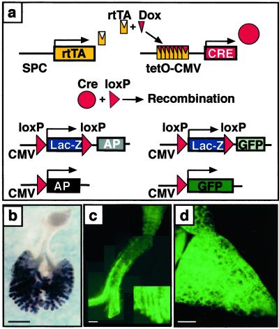
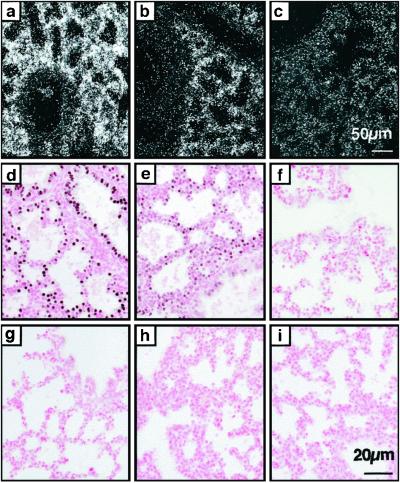
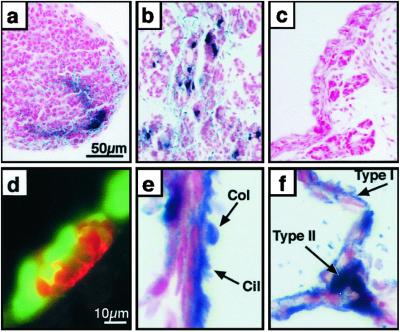
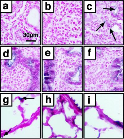
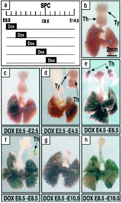
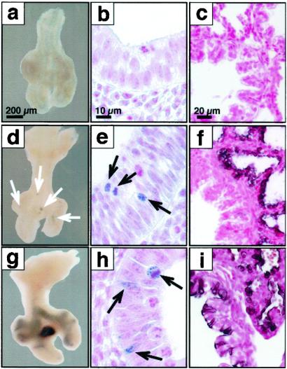
To establish the timing of lineage restriction among endodermal derivatives, we developed a method to label permanently subsets of lung precursor cells at defined times during development by using Cre recombinase to activate floxed alkaline phosphatase or green fluorescent protein genes under control of doxycycline-dependent surfactant protein C promoter. Extensive or complete labeling of peripheral lung, thyroid, and thymic epithelia, but not trachea, bronchi, or gastrointestinal tract occurred when mice were exposed to doxycycline from embryonic day (E) 4.5 to E6.5. Nonoverlapping cell lineages of conducting airways (trachea and bronchi), as distinct from those of peripheral airways (bronchioles, acini, and alveoli), were established well before formation of the definitive lung buds at E9-9.5. At E11.5, the labeled precursors of peripheral lung were restricted to relatively few cells along the bronchial tubes and clusters in bronchial tips and lateral buds. Thereafter, these cells underwent marked expansion to form the entire gas-exchange region in the lung. This study demonstrates early restriction of endodermal progenitor cells forming peripheral as compared with proximal airways, identifies distinct cell lineages in conducting airways, and distinguishes neuroepithelial and tracheal-bronchial gland cell lineages from those lining peripheral regions of the lung. This system for conditional gene addition or deletion is useful for the study of lung morphogenesis and gene function in vivo, and identifies progenitor cells that may serve as useful targets for cell or gene replacement for pulmonary disorders.


Figures

Fig 1.
Fig 1.
Permanent cell labeling by doxycycline-inducible recombination in triple-transgenic mice. (a) Triple-transgenic mice were produced in which doxycycline activates rtTA expressed under control of the SP-C promoter. The rtTA then activates Cre that excises and religates the target gene, inactivating β-galactosidase (lacZ), and permanently labeling the cell by activating AP or GFP in the ZAP or ZEG mice, respectively. Dams were treated with doxycycline from E6.5 to day of killing. (b) Whole mount AP staining on an E13.5 lung from an SP-C-rtTA/tetO-Cre/ZAP embryo. Most intrapulmonary epithelial cells and some extrapulmonary airway cells stained for AP. (c) GFP fluorescence in trachea from an SP-C-rtTA/tetO-Cre/ZEG transgenic mouse on PN7. GFP-positive cells are organized in a linear fashion along the trachea and numbers of labeled cells increased from proximal to peripheral extrapulmonary airways (lower right corner, magnification, ×5). (d) Widespread GFP fluorescence was seen in lung parenchyma from an SP-C-rtTA/tetO-Cre/ZEG mouse on PN7. (Bars = 1 mm.)
Fig 2.
Fig 2.
Decreased Cre mRNA and protein after removal of doxycycline. Dams were treated for 48 h with doxycycline in the food. In situ hybridization for Cre mRNA (ac) and immunohistochemistry for Cre protein (di) was performed on lung sections from embryos on E18.5, whereas on doxycycline (a, d, and g), 48 h (b, e, and h) and 96 h (c, f, and i) after removal from doxycycline, triple transgenic embryos (af), and double-transgenic embryos (gi). (ac) Cre-mRNA was detected throughout the respiratory epithelium, whereas on doxycycline, decreased after 48 h and was not different from nontransgenic controls (data not shown) 96 h after removal from doxycycline. [Bar = 50 μm (ac).] (df) Whereas on doxycycline, Cre protein was readily detected in nuclei of most pulmonary epithelial cells, Cre protein was reduced 48 h after cessation of doxycycline and absent after 96 h. (gi) In embryos lacking the SP-C-rtTA transgene (double-transgenic Cre/ZAP), Cre protein was not detected on or off doxycycline. [Bar = 20 μm (di).]
Fig 3.
Fig 3.
Cre-mediated recombination in diverse epithelial cell types: thymus, thyroid, and peripheral lung. Pregnant dams were treated with doxycycline from E0.5 to E14.5 (a) or E0.5 to PN7 (bf). AP staining (ac, e, and f) was performed on paraffin sections counterstained with nuclear fast red. (a) AP staining of cells in the apical region of the thymus. (b) Subsets of thyroid cells expressed AP. (c) Tracheal-bronchial glands do not stain for AP. (d) Immunohistochemistry for CGRP (red) stained neuroendocrine bodies (NEBs) that did not coexpress GFP (green). (e) Most ciliated (cil) and nonciliated Clara (col) cells in the bronchioles stain for AP. (f) Staining in alveolar type I (type I) and type II (type II) cells was detected. [Bar = 50 μm (ac) and 10 μm (df).]
Fig 4.
Fig 4.
Cre-mediated recombination in the embryonic and postnatal lung. Lung sections were obtained from mice killed at E14.5 (a and d), E15.5 (b and e), E16.5 (c and f), and PN 21 (gi), and stained for AP with nuclear fast red counterstain. Recombination in the absence of doxycycline is rare (arrows) and can be seen only after E15.5 (ac and g). Cell labeling was widespread after treatment of the dam with doxycycline from E6.5 to E14.5, E15.5, and E16.5 (df). Rare labeling was observed when exposed to doxycycline from E18.5 to PN9 (h) or PN2 to PN9 (i). [Bar = 30 μm (ai).]
Fig 5.
Fig 5.
Temporal specificity of recombination. Dams were treated with doxycycline for 48 h and labeling assessed at E14.5 by whole-mount AP staining. A diagram of the treatment protocols used during embryonic development is shown (a). E0.5 is 12 h after fertilization as determined by detection of a vaginal plug. Whole-mount AP staining of lungs from triple-transgenic mice was assessed on E14.5 to determine the extent of recombination (bh). Single-transgenic control littermate (b). Partial lobar labeling in the peripheral lung after doxycycline from E0.5 to E2.5 (c). Extensive labeling of intrapulmonary tissue with variable staining in the thymus and thyroid seen among littermates (d). Labeling of most intrapulmonary epithelial cells. Staining was observed in the thymus of all mice but not in the thyroid (e). Labeling in the peripheral lung was found in all mice; recombination in the thymus was found frequently but not in all mice (f). Labeling of extrapulmonary airways was never observed from E0.5to E8.5 (cf). From E8.5 to E10.5, labeling of intrapulmonary epithelial cells was widespread or focal with variation seen among littermates; subsets of cells in extrapulmonary airways were labeled, the numbers of labeled cells being more abundant in the periphery, and labeling was not seen in thymus or thyroid (g and h). Extensive, but not complete labeling of intrapulmonary epithelial cells was observed in association with labeling of some cells in the extrapulmonary airways (h). (Bar = 2 mm.) Th, thymus; Ty, thyroid.
Fig 6.
Fig 6.
Progenitors of the peripheral lung are highly restricted at E11.5. Lungs were obtained at E11.5 (a, b, d, e, g, and h), at E18.5 (c, f, and i), from control mice (ac) or triple-transgenic mice SP-C-rtTA/(tetO)Cre/Zap (d, f, g, and i) and SP-C-rtTA/(tetO)Cre/ZEG (e and h) after doxycycline treatment from E6.5 to E8.5 (df) or E8.5 to E10.5 (gi). No AP or GFP staining was seen in controls. At E11.5, AP-labeled cells seen after treatment with doxycycline from E6.5 to E8.5 were highly restricted to small clusters along the bronchial tubes and to the tips of the lobar bronchi (d). At higher magnification, small clusters of GFP-immunostaining cells were noted at these locations (e). This same period of doxycycline exposure resulted in nearly complete labeling of peripheral lung with exclusion of large conducting and extrapulmonary airways at E18.5 (f). After doxycycline exposure, from E8.5 to E10.5, more extensive AP (g) or GFP (h) was seen in proximal and peripheral lung tubules at E11.5. Consistent with the more widespread labeling of extrapulmonary conducting airways seen at E18.5, patchy labeling of the peripheral lung parenchyma was also noted at E18.5 (i). [Bars = 200 μm, (a, d, g), 10 μm, (b, e, h), and 20 μm (c, f, i).]

References

    1. Wells J. M. & Melton, D. A. (1999) Annu. Rev. Cell. Dev. Biol. 15, 393-410. - PubMed
    1. Wells J. M. & Melton, D. A. (2000) Development (Cambridge, U.K.) 127, 1563-1572. - PubMed
    1. Lawson K. A., Meneses, J. J. & Pedersen, R. A. (1986) Dev. Biol. 115, 325-339. - PubMed
    1. Perl A.-K. T. & Whitsett, J. A. (1999) Clin. Genet. 56, 14-27. - PubMed
    1. Warburton D., Schwarz, M., Tefft, D., Flores-Delgado, G., Anderson, K. D. & Cardoso, W. V. (2000) Mech. Dev. 92, 55-81. - PubMed

Publication types

MeSH terms

LinkOut - more resources