. 2002 Aug 6;99(16):10843-8.
doi: 10.1073/pnas.162228299. Epub 2002 Jul 26.

Clusterin promotes amyloid plaque formation and is critical for neuritic toxicity in a mouse model of Alzheimer's disease

Affiliations

Clusterin promotes amyloid plaque formation and is critical for neuritic toxicity in a mouse model of Alzheimer's disease

Ronald B DeMattos et al. Proc Natl Acad Sci U S A. .

Abstract

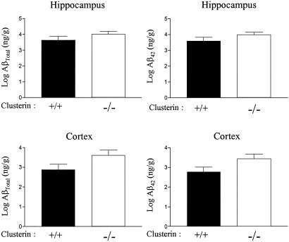
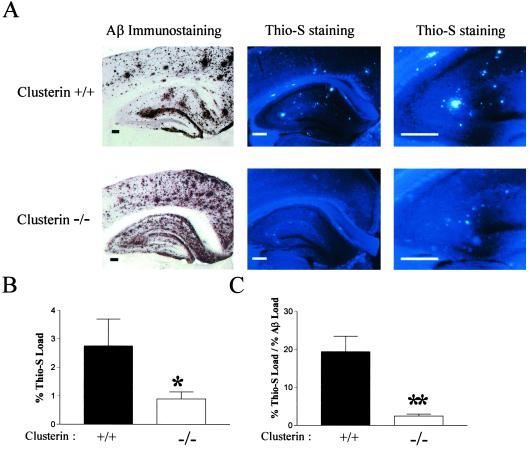
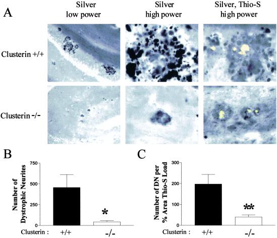
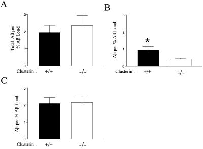
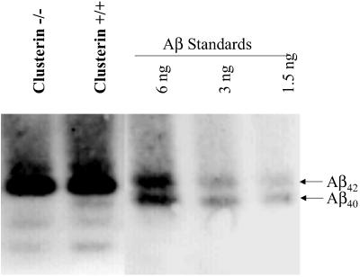
Studies have shown that clusterin (also called apolipoprotein J) can influence the structure and toxicity of amyloid-beta (Abeta) in vitro. To determine whether endogenous clusterin plays a role in influencing Abeta deposition, structure, and toxicity in vivo, we bred PDAPP mice, a transgenic mouse model of Alzheimer's disease, to clusterin(-/-) mice. By 12 months of age, PDAPP, clusterin(-/-) mice had similar levels of brain Abeta deposition as did PDAPP, clusterin(+/+) mice. Although Abeta deposition was similar, PDAPP, clusterin(-/-) mice had significantly fewer fibrillar Abeta (amyloid) deposits than PDAPP mice expressing clusterin. In the absence of clusterin, neuritic dystrophy associated with the deposited amyloid was markedly reduced, resulting in a dissociation between fibrillar amyloid formation and neuritic dystrophy. These findings demonstrate that clusterin markedly influences Abeta structure and neuritic toxicity in vivo and is likely to play an important role in Alzheimer's disease pathogenesis.


Figures

Fig 1.
Fig 1.
Clusterin expression does not alter the mass of deposited Aβ in PDAPP mice. Levels of AβTotal or Aβ42 as assessed by ELISA on guanidine extracts from hippocampus and cortex did not reveal significant differences between 12-month-old PDAPP+/+, clusterin+/+ (n = 13) versus PDAPP+/+, clusterin−/− mice (n = 14). Data reported are means ± SEM.
Fig 2.
Fig 2.
Clusterin facilitates the conversion of Aβ into amyloid in vivo. The 12-month-old PDAPP+/+, clusterin+/+ and PDAPP+/+, clusterin−/− mice containing Aβ-immunoreactive deposits were analyzed for the presence of thioflavine-S (Thio-S)-positive plaques (amyloid). (A) Serial brain sections were either immunostained with either a polyclonal antibody against Aβ (Left) or with the dye thioflavine-S (Center and Right). (Scale bars: Left, 500 μm; Center, 250 μm; Right, 125 μm.) (B) PDAPP+/+, clusterin−/− mice (n = 15) had significantly less hippocampal thioflavine-S load than littermate PDAPP+/+, clusterin+/+ mice (n = 13). *, P = 0.05. (C) The percentage of deposited Aβ that was thioflavine-S-positive (fibrillar) was significantly decreased in PDAPP+/+, clusterin−/− mice. **, P < 0.0001. Data in B and C are means ± SEM.
Fig 3.
Fig 3.
Dissociation between amyloid plaques and neurite toxicity in PDAPP+/+, clusterin−/− mice. (A) Brain sections from 12-month-old PDAPP+/+, clusterin+/+ and PDAPP+/+, clusterin−/− mice were labeled with the de Olmos silver stain with or without thioflavine-S (Thio-S) to identify the neuritic dystrophy associated with the fibrillar amyloid. Vast numbers of dystrophic neurites (DN) were observed in the locale of thioflavine-S-positive deposits in PDAPP+/+, clusterin+/+ mice (Upper) at low and high power. Little neuritic dystrophy surrounded thioflavine-S-positive deposits in the PDAPP+/+, clusterin−/− mice (Lower). (B) PDAPP+/+, clusterin−/− mice had significantly fewer dystrophic neurites (mean ± SEM: 42.9 ± 13.8, n = 15) in three equally spaced sections than PDAPP+/+, clusterin+/+ mice (456.6 ± 155.2, n = 13). *, P = 0.0083. (C) The number of dystrophic neurites normalized to the percent area of the hippocampus covered by thioflavine-S was significantly decreased (5-fold) in PDAPP+/+, clusterin−/− mice (mean ± SEM: 40.0 ± 10.1, n = 15) compared with the PDAPP+/+, clusterin+/+ mice (197.3 ± 45.8, n = 13). **, P = 0.0014.
Fig 4.
Fig 4.
Clusterin expression alters the soluble pool of brain Aβ. (A) Carbonate-soluble hippocampal extracts from 12-month-old PDAPP+/+, clusterin+/+ (n = 13) and PDAPP+/+, clusterin−/− (n = 15) mice had similar levels of AβTotal when assayed by ELISA under denaturing conditions. The AβTotal concentrations (nanograms per milligram of protein) were normalized to the percent Aβ load to correct for the variability in deposition between animals. (B) When the carbonate extracts were analyzed under nondenaturing conditions with the same ELISA, mice expressing clusterin had a significant 2-fold increase in the soluble pool of AβTotal. *, P = 0.01. (C) Carbonate extracts analyzed under nondenaturing conditions with an ELISA by using the same capture and detecting antibody (resulting in an assay that should be specific for oligomeric forms of Aβ) did not detect a significant difference between PDAPP+/+, clusterin+/+ and PDAPP+/+, clusterin−/− mice. Data in B and C are means ± SEM.
Fig 5.
Fig 5.
The subtle difference in the Aβ peptide composition of the soluble pool of brain Aβ is revealed by acid-urea polyacrylamide gels. The Aβ peptide composition of the carbonate-soluble hippocampal extracts from 12-month-old PDAPP+/+, clusterin+/+ and PDAPP+/+, clusterin−/− mice was determined by acid-urea gel analysis (n = 4 per group). Human Aβ40 and Aβ42 synthetic peptides are used for mass and migration comparisons. Soluble extracts analyzed under the completely denaturing conditions demonstrate that human Aβ42 is the predominant Aβ peptide present. Human Aβ40 was readily detectable in extracts from all PDAPP+/+, clusterin+/+ mice examined, whereas human Aβ40 could not be detected in PDAPP+/+, clusterin−/− extracts.

References

    1. Selkoe D. J. (2001) Physiol. Rev. 81, 741-766. - PubMed
    1. Sisodia S. S. (1999) J. Clin. Invest. 104, 1169-1170. - PMC - PubMed
    1. Van Nostrand W. E., Melchor, J. P., Cho, H. S., Greenberg, S. M. & Rebeck, G. W. (2001) J. Biol. Chem. 276, 32860-32866. - PubMed
    1. Nilsberth C., Westlind-Danielsson, A., Eckman, C. B., Condron, M. M., Axelman, K., Forsell, C., Stenh, C., Luthman, J., Teplow, D. B., Younkin, S. G., et al. (2001) Nat. Neurosci. 4, 887-893. - PubMed
    1. Strittmatter W. J., Saunders, A. M., Schmechel, D., Pericak-Vance, M., Enghild, J., Salvesen, G. S. & Roses, A. D. (1993) Proc. Natl. Acad. Sci. USA 90, 1977-1981. - PMC - PubMed

Publication types

MeSH terms