

doi: 10.1086/342193.
Epub 2002 Jul 24.
Mutations in a novel gene, TMIE, are associated with hearing loss linked to the DFNB6 locus
Affiliations
- PMID: 12145746
- PMCID: PMC379198
- DOI: 10.1086/342193
Item in Clipboard
Mutations in a novel gene, TMIE, are associated with hearing loss linked to the DFNB6 locus
Am J Hum Genet.
2002 Sep.
Abstract
We have identified five different homozygous recessive mutations in a novel gene, TMIE (transmembrane inner ear expressed gene), in affected members of consanguineous families segregating severe-to-profound prelingual deafness, consistent with linkage to DFNB6. The mutations include an insertion, a deletion, and three missense mutations, and they indicate that loss of function of TMIE causes hearing loss in humans. TMIE encodes a protein with 156 amino acids and exhibits no significant nucleotide or deduced amino acid sequence similarity to any other gene.
Figures
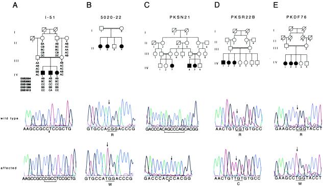
Families showing linkage to DFNB6 and TMIE mutation analyses. A, Family I-51, from India, defined the minimum interval for DFNB6 as being between markers D3S3582 and D3S1613. Sequence chromatograms from a noncarrier (II:4) and a deaf individual (IV:2) show the wild-type sequence, the place of insertion (arrowhead), and the insertion of CGCC (bracketed) in exon 2, respectively. B, 250C→T transition in exon 3 in an affected individual (II:1) from family 5020-22, compared with the wild-type sequence. C, The chromatograms show sequence of the intron 1–exon 2 boundary in PKSN21, from a noncarrier and an affected individual with the deletion of seven bases (bracketed in wild type). The site of the deletion is indicated by an arrowhead, along with an insertion of cytosine (arrow). D, The sequence traces of a wild type and an affected individual (IV:2) in family PKSR22B with the 241C→T transition (arrows). E, Chromatograms from an unaffected control individual and an affected individual (IV:1) from family PKDF76, showing the 274C→T transition (arrows).
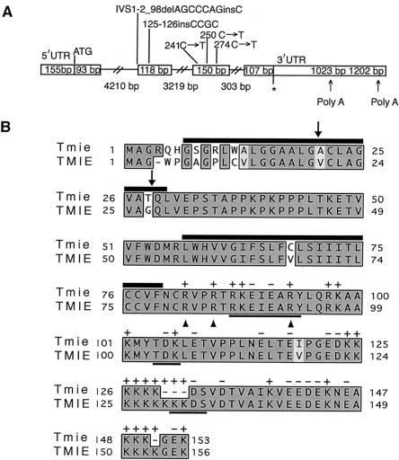
Diagrammatic representations of TMIE and TMIE. A, The boxed regions depict exons of TMIE. Slashed lines represent introns. The length of each exon and intron is given in base pairs (bp), within boxes (for exons) and below slashed lines (for introns). “ATG” denotes the initiation codon, and the asterisk (*) marks the stop codon. Mutations found in the five families showing linkage to DFNB6 are shown above the respective exons. We obtained a 1023-bp 3′ UTR followed by a poly A tail. BLAST analysis of the TMIE cDNA sequence identified ESTs corresponding to a 1202-bp 3′ UTR because of utilization of a different polyadenylation signal. B, Clustal W alignment of deduced amino acid sequences of Tmie and TMIE. The shared amino acid identity between Tmie (mouse) and TMIE (human) is indicated by shaded boxes: dark gray for absolute identity and light gray for conservative changes. Dashes (—) show gaps in the alignment. TMIE has two potential sites of signal peptide cleavage (arrows), and two predicted transmembrane regions (black bars on top of amino acid residues). Cleavage of the signal peptide would result in a protein with an extracellular amino terminus, one transmembrane segment, and an intracellular carboxy terminus. The C-terminus has many positively charged amino acids interspersed with negatively charged residues (charge indicated on top of each residue) and three potential protein kinase phosphorylation sites (bold underlines). Arrowheads indicate arginine residues in TMIE mutated in affected individuals in three families linked to DFNB6.
Similar articles
-
Mutations in a novel gene with transmembrane domains underlie Usher syndrome type 3.Am J Hum Genet. 2001 Oct;69(4):673-84. doi: 10.1086/323610. Epub 2001 Aug 27. Am J Hum Genet. 2001. PMID: 11524702 Free PMC article.
-
Homozygous mutations in Pakistani consanguineous families with prelingual nonsyndromic hearing loss.Mol Biol Rep. 2020 Dec;47(12):9979-9985. doi: 10.1007/s11033-020-06037-7. Epub 2020 Dec 2. Mol Biol Rep. 2020. PMID: 33269433
-
Mutation of the novel gene Tmie results in sensory cell defects in the inner ear of spinner, a mouse model of human hearing loss DFNB6.Hum Mol Genet. 2002 Aug 1;11(16):1887-98. doi: 10.1093/hmg/11.16.1887. Hum Mol Genet. 2002. PMID: 12140191
-
Mutations of human TMHS cause recessively inherited non-syndromic hearing loss.J Med Genet. 2006 Aug;43(8):634-40. doi: 10.1136/jmg.2005.039834. Epub 2006 Feb 3. J Med Genet. 2006. PMID: 16459341 Free PMC article.
-
Analysis of TMIE gene mutations including the first large deletion of exon 1 with autosomal recessive non-syndromic deafness.BMC Med Genomics. 2022 Jun 16;15(1):133. doi: 10.1186/s12920-022-01287-9. BMC Med Genomics. 2022. PMID: 35710363 Free PMC article.
Cited by
-
Molecular Identity of the Mechanotransduction Channel in Hair Cells: Not Quiet There Yet.J Neurosci. 2016 Oct 26;36(43):10927-10934. doi: 10.1523/JNEUROSCI.1149-16.2016. J Neurosci. 2016. PMID: 27798175 Free PMC article.
-
Extending the knowledge in histochemistry and cell biology.Histochem Cell Biol. 2010 Jan;133(1):1-40. doi: 10.1007/s00418-009-0665-2. Epub 2009 Nov 28. Histochem Cell Biol. 2010. PMID: 19946696 Review.
-
Mechanically Activated Ion Channels.Neuron. 2015 Sep 23;87(6):1162-1179. doi: 10.1016/j.neuron.2015.08.032. Neuron. 2015. PMID: 26402601 Free PMC article. Review.
-
USH1H, a novel locus for type I Usher syndrome, maps to chromosome 15q22-23.Clin Genet. 2009 Jan;75(1):86-91. doi: 10.1111/j.1399-0004.2008.01038.x. Epub 2008 May 25. Clin Genet. 2009. PMID: 18505454 Free PMC article.
-
Global genetic insight contributed by consanguineous Pakistani families segregating hearing loss.Hum Mutat. 2019 Jan;40(1):53-72. doi: 10.1002/humu.23666. Epub 2018 Nov 18. Hum Mutat. 2019. PMID: 30303587 Free PMC article.
References
Electronic-Database Information
-
- Connexins and Deafness Homepage, http://www.crg.es/deafness/ (for population differences in carrier rates and deafness due to CX26 mutations)
-
- GenBank Overview, http://www.ncbi.nlm.nih.gov/Genbank/GenbankOverview.html (for human TMIE, [accession number AY081842] and D. melanogaster hypothetical protein [CG15130; accession number AAF53893.1])
-
- Genscan, http://genes.mit.edu/GENSCAN.html (for cryptic splice-site prediction)
-
- Hereditary Hearing Loss Homepage, http://dnalab-www.uia.ac.be/dnalab/hhh/ (for a list of all known genes causing hearing loss)
-
- NCBI BLAST home page, http://www.ncbi.nlm.nih.gov/BLAST/ (for TMIE homologs)
References
-
- Adams MD, Celniker SE, Holt RA, Evans CA, Gocayne JD, Amanatides PG, Scherer SE, et al (2000) The genome sequence of Drosophila melanogaster. Science 287:2185–2195 - PubMed
-
- Ahmed ZM, Smith TN, Riazuddin S, Makishima T, Ghosh M, Bokhari S, Menon SNP, Deshmukh D, Griffith AJ, Riazuddin S, Friedman TB, Wilcox ER (2002) Nonsyndromic recessive deafness DFNB18 and Usher syndrome type IC are allelic mutations of USH1C. Hum Genet 110:527–531 - PubMed
-
- Fukushima K, Ramesh A, Srisailapathy CR, Ni L, Wayne S, O'Neill ME, Van Camp G, Coucke P, Jain P, Wilcox ER, Smith SD, Kenyon JB, Zbar RIS, Smith RJH (1995) An autosomal recessive nonsyndromic form of sensorineural hearing loss maps to 3p-DFNB6. Genome Res 5:305–308 - PubMed
-
- Hmani M, Ghorbel A, Boulila-Elgaied A, Ben Zina Z, Kammoun W, Drira M, Chaabouni M, Petit C, Ayadi H (1999) A novel locus for Usher syndrome type II, USH2B, maps to chromosome 3 at p23-24.2. Eur J Hum Genet 7:363–367 - PubMed
Publication types
MeSH terms
Substances
Associated data
- Actions
Grants and funding
LinkOut - more resources
Full Text Sources
Other Literature Sources
Medical
Molecular Biology Databases
