

The peptidyl-prolyl isomerase motif is lacking in PmpA, the PrsA-like protein involved in the secretion machinery of Lactococcus lactis
- PMID: 12147493
- PMCID: PMC124044
- DOI: 10.1128/AEM.68.8.3932-3942.2002
The peptidyl-prolyl isomerase motif is lacking in PmpA, the PrsA-like protein involved in the secretion machinery of Lactococcus lactis
Abstract
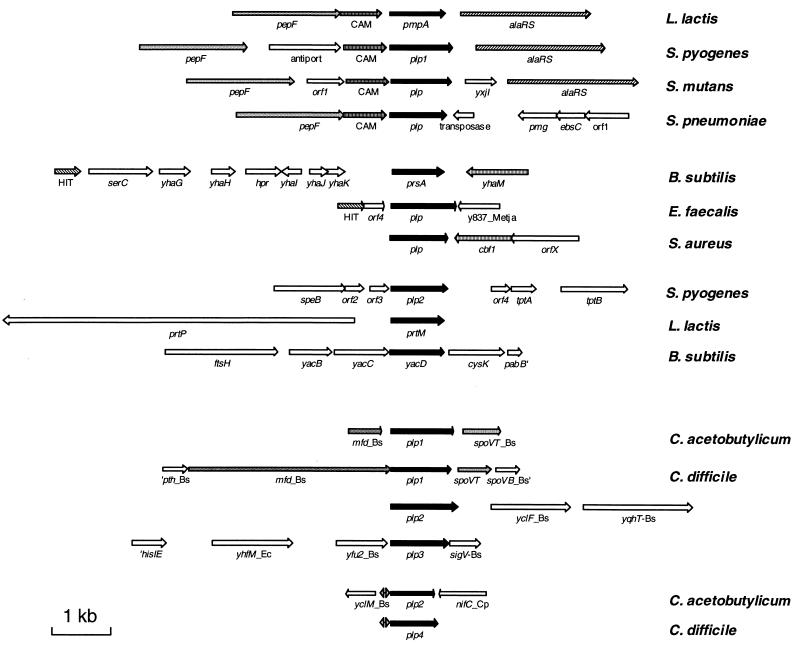
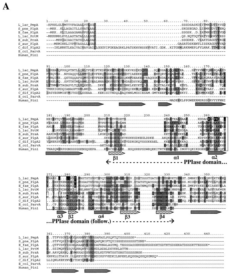
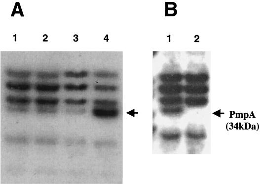
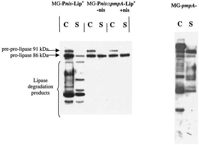
The prsA-like gene from Lactococcus lactis encoding its single homologue to PrsA, an essential protein triggering the folding of secreted proteins in Bacillus subtilis, was characterized. This gene, annotated pmpA, encodes a lipoprotein of 309 residues whose expression is increased 7- to 10-fold when the source of nitrogen is limited. A slight increase in the expression of the PrsA-like protein (PLP) in L. lactis removed the degradation products previously observed with the Staphylococcus hyicus lipase used as a model secreted protein. This shows that PmpA either triggers the folding of the secreted lipase or activates its degradation by the cell surface protease HtrA. Unlike the case for B. subtilis, the inactivation of the gene encoding PmpA reduced only slightly the growth rate of L. lactis in standard conditions. However, it almost stopped its growth when the lipase was overexpressed in the presence of salt in the medium. Like PrsA of B. subtilis and PrtM of L. lactis, the L. lactis PmpA protein could thus have a foldase activity that facilitates protein secretion. These proteins belong to the third family of peptidyl-prolyl cis/trans-isomerases (PPIases) for which parvulin is the prototype. Almost all PLP from gram-positive bacteria contain a domain with the PPIase signature. An exception to this situation was found only in Streptococcaceae, the family to which L. lactis belongs. PLP from Streptococcus pneumoniae and Enterococcus faecalis possess this signature, but those of L. lactis, Streptococcus pyogenes, and Streptococcus mutans do not. However, secondary structure predictions suggest that the folding of PLP is conserved over the entire length of the proteins, including the unconserved signature region. The activity associated with the expression of PmpA in L. lactis and these genomic data show that either the PPIase motif is not necessary for PPIase activity or, more likely, PmpA foldase activity does not necessarily require PPIase activity.
Figures

Similar articles
-
Structure-function analysis of PrsA reveals roles for the parvulin-like and flanking N- and C-terminal domains in protein folding and secretion in Bacillus subtilis.J Biol Chem. 2004 Apr 30;279(18):19302-14. doi: 10.1074/jbc.M400861200. Epub 2004 Feb 19. J Biol Chem. 2004. PMID: 14976191
-
Structural analysis of extracellular ATP-independent chaperones of streptococcal species and protein substrate interactions.mSphere. 2025 Feb 25;10(2):e0107824. doi: 10.1128/msphere.01078-24. Epub 2025 Jan 29. mSphere. 2025. PMID: 39878509 Free PMC article.
-
Characterisation of SEQ0694 (PrsA/PrtM) of Streptococcus equi as a functional peptidyl-prolyl isomerase affecting multiple secreted protein substrates.Mol Biosyst. 2015 Dec;11(12):3279-86. doi: 10.1039/c5mb00543d. Mol Biosyst. 2015. PMID: 26466087
-
Archaeal peptidyl prolyl cis-trans isomerases (PPIases) update 2004.Front Biosci. 2004 May 1;9:1680-720. doi: 10.2741/1361. Front Biosci. 2004. PMID: 14977579 Review.
-
Archaeal peptidyl prolyl cis-trans isomerases (PPIases).Front Biosci. 2000 Sep 1;5:D821-36. doi: 10.2741/maruyama. Front Biosci. 2000. PMID: 10966874 Review.
Cited by
-
Identification of Conserved and Species-Specific Functions of the Listeria monocytogenes PrsA2 Secretion Chaperone.Infect Immun. 2015 Oct;83(10):4028-41. doi: 10.1128/IAI.00504-15. Epub 2015 Jul 27. Infect Immun. 2015. PMID: 26216425 Free PMC article.
-
Phenotypic characterization of the foldase homologue PrsA in Streptococcus mutans.Mol Oral Microbiol. 2013 Apr;28(2):154-65. doi: 10.1111/omi.12014. Epub 2012 Dec 13. Mol Oral Microbiol. 2013. PMID: 23241367 Free PMC article.
-
PpiA, a surface PPIase of the cyclophilin family in Lactococcus lactis.PLoS One. 2012;7(3):e33516. doi: 10.1371/journal.pone.0033516. Epub 2012 Mar 19. PLoS One. 2012. PMID: 22442694 Free PMC article.
-
Dimeric Structure of the Bacterial Extracellular Foldase PrsA.J Biol Chem. 2015 Feb 6;290(6):3278-92. doi: 10.1074/jbc.M114.622910. Epub 2014 Dec 17. J Biol Chem. 2015. PMID: 25525259 Free PMC article.
-
Listeria monocytogenes virulence factor secretion: don't leave the cell without a chaperone.Front Cell Infect Microbiol. 2014 Feb 12;4:13. doi: 10.3389/fcimb.2014.00013. eCollection 2014. Front Cell Infect Microbiol. 2014. PMID: 24575392 Free PMC article. Review.
References
-
- Arie, J. P., N. Sassoon, and J. M. Betton. 2001. Chaperone function of FkpA, a heat shock prolyl isomerase, in the periplasm of Escherichia coli. Mol. Microbiol. 39:199-210. - PubMed
-
- Arnau, J., E. Hjerl-Hansen, and H. Israelsen. 1997. Heterologous gene expression of bovine plasmin in Lactococcus lactis. Appl. Microbiol. Biotechnol. 48:331-338. - PubMed
Publication types
MeSH terms
Substances
LinkOut - more resources
Full Text Sources
Other Literature Sources
Molecular Biology Databases
Research Materials
