

Review
doi: 10.1083/jcb.200205034.
Epub 2002 Jul 29.
Mycobacterium and the coat of many lipids
Affiliations
- PMID: 12147678
- PMCID: PMC2173834
- DOI: 10.1083/jcb.200205034
Item in Clipboard
Review
Mycobacterium and the coat of many lipids
J Cell Biol.
.
Abstract
Pathogenic Mycobacterium reside inside vacuoles in their host macrophages. These vacuoles fail to fuse with lysosomes yet interact with early endosomes. Glycoconjugates released by the intracellular bacilli traffic through the host cell and are released through exocytosis. These molecules represent both antigens for immune recognition and modulators of immune function. The molecules play key roles in the induction and maintenance of the granuloma, a tissue response that limits bacterial spread yet ensures persistence of the infection.
Figures

Electron micrograph of an alveolar macrophage isolated by broncholavage from a tuberculosis patient in Malawi. The cell is heavily loaded with bacteria that reside within vacuoles. The vacuoles vary both in size and in the number of bacteria. This degree of heterogeneity in vacuole morphology is not observed in infections in culture.
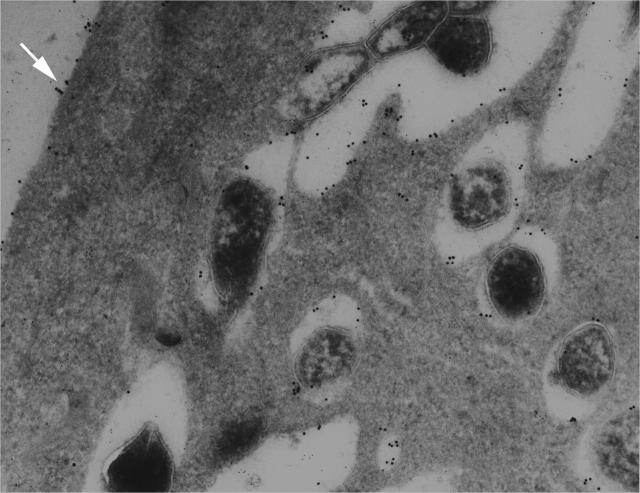
Bone marrow–derived macrophages infected 72 h previously with M. tuberculosis, incubated in 10 μg/ml biotinylated cholera toxin B for 15 min, washed, and placed in prewarmed medium for a 45 min chase period. The section was probed with streptavidin/antistreptavidin (anti–rabbit IgG 18 nm gold). Cholera toxin B can be detected in all of the bacterial vacuoles visible in this field, demonstrating the continued interaction between the host cell plasmalemma and the vacuoles harboring the bacilli. Reproduced from Russell et al. (1996).
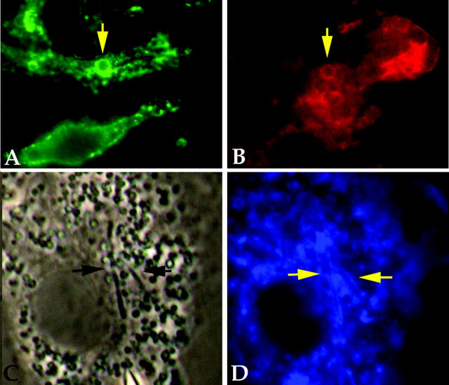
Immunofluorescent analysis of macrophage phagosomes formed around IgG beads (A/B) and
M. tuberculosis
(C/D). Fluorescent analysis of macrophage after uptake of IgG-coated beads (A and B) and infection with M. tuberculosis (C and D). Macrophages fixed 15 min after internalization of IgG-coated beads (arrows) probed with antibodies against LAMP1 (A) and coronin I (B). The labeling pattern shows strong colocalization of both LAMP1 and coronin I around the early phagosome. C and D shows phase–contrast and fluorescence micrographs, respectively, of macrophages fixed and probed with filipin after overnight infection with M. tuberculosis. Filipin associates directly with cholesterol and shows the distribution of the lipid in the cell. There is a clear halo around the intracellular bacteria (arrows).
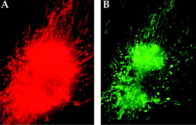
Release of labeled mycobacterial cell wall lipids into the macrophage endocytic network. (A) A live bone marrow–derived macrophage infected for 24 h with Texas red hydrazide–labeled BCG were analyzed by fluorescence microscopy, with striking release of Texas red label from the bacterial phagosome. (B) Infected macrophages were incubated with dextran-fluorescein for 1 h followed by a chase period of 3 h. Fluorescent label (Texas red) released from the bacteria permeated the host macrophage and colocalized with dextran-fluorescein, revealing the accessibility of mycobacterial constituents to endocytic compartments. Reproduced from Beatty et al. (2000).
References
-
- Beatty, W.B., E.R. Rhoades, H.J. Ullrich, D. Chatterjee, and D.G. Russell. 2000. Trafficking and release of mycobacterial lipids from infected macrophages. Traffic. 1:235–247. - PubMed
-
- Beatty, W.L., H.J. Ullrich, and D.G. Russell. 2001. Mycobacterial surface moieties are released from infected macrophages by a constitutive exocytic event. Eur. J. Cell Biol. 80:31–40. - PubMed
-
- Besra, G.S., and D. Chatterjee. 1994. Lipids and carbohydrates of Mycobacterium tuberculosis. Tuberculosis: Pathogenesis, Protection, and Control. B.R. Bloom, editor. ASM Press, Washington, D.C. 285–306.
-
- Brightbill, H.D., D.H. Libraty, S.R. Krutzik, R.B. Yang, J.T. Belisle, J.R. Bleharski, M. Maitland, M.V. Norgard, S.E. Plevy, S.T. Smale, et al. 1999. Host defense mechanisms triggered by microbial lipoproteins through toll-like receptors. Science. 285:732–736. - PubMed
Publication types
MeSH terms
Substances
Grants and funding
LinkOut - more resources
Full Text Sources
Other Literature Sources
Medical
