

Review
doi: 10.1083/jcb.200205110.
Epub 2002 Jul 29.
Lysosomes and the plasma membrane: trypanosomes reveal a secret relationship
Affiliations
- PMID: 12147679
- PMCID: PMC2173812
- DOI: 10.1083/jcb.200205110
Item in Clipboard
Review
Lysosomes and the plasma membrane: trypanosomes reveal a secret relationship
J Cell Biol.
.
Abstract
Studies of the cell invasion mechanism of the parasite Trypanosoma cruzi led to a series of novel findings, which revealed a previously unsuspected ability of conventional lysosomes to fuse with the plasma membrane. This regulated exocytic process, previously regarded mostly as a specialization of certain cell types, was recently shown to play an important role in the mechanism by which cells reseal their plasma membrane after injury.
Figures
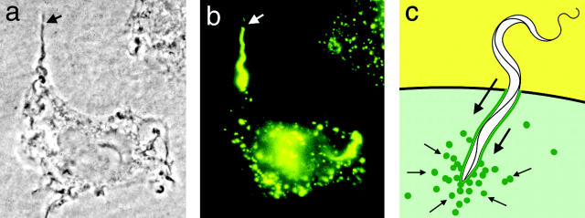
The lysosome-mediated cell invasion mechanism of T. cruzi. (a) Phase-contrast image of a trypomastigote in the process of entering a HeLa cell. The arrow points to the extracellular portion of the parasite. (b) Immunofluorescence image of the same cell shown in panel a stained with antibodies against human Lamp-1. The arrow points to the extracellular portion of the parasite, not yet surrounded by Lamp-1–containing membranes. (c) Diagram of the process originating the trypomastigote-containing intracellular vacuole. The green line represents lysosomal membranes that are gradually incorporated into the vacuole, the small arrows indicate the direction of lysosome movement, and the large arrows indicate the direction of parasite movement.
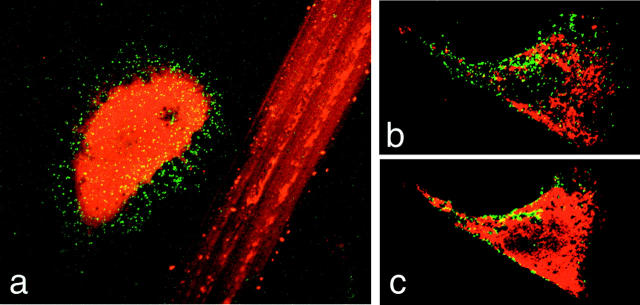
Exposure of the lumenal domain of Lamp-1 on the surface of wounded cells. (a) A wounded NRK cell, containing cytosolic Texas red–dextran, is shown next to the track left on the coverslip by scratching the monolayer. (b and c) Sequential confocal Z-sections through the bottom (b) and middle (c) of a wounded 3T3 fibroblast. In all images, the green/yellow punctate staining corresponds to the lumenal epitope of Lamp-1, recognized by a monoclonal antibody on the surface of nonpermeabilized cells.
References
-
- Andrews, N.W., and M.B. Whitlow. 1989. Secretion by Trypanosoma cruzi of a hemolysin active at low pH. Mol. Biochem. Parasitol. 33:249–256. - PubMed
-
- Ashino, Y., X. Ying, L.G. Dobbs, and J. Bhattacharya. 2000. [Ca(2+)](i) oscillations regulate type II cell exocytosis in the pulmonary alveolus. Am. J. Physiol. Lung Cell. Mol. Physiol. 279:L5–L13. - PubMed
-
- Baetz, K., S. Isaaz, and G.M. Griffiths. 1995. Loss of cytotoxic T lymphocyte function in Chediak-Higashi syndrome arises from a secretory defect that prevents lytic granule exocytosis. J. Immunol. 154:6122–6131. - PubMed
-
- Bakker, A.C., P. Webster, W.A. Jacob, and N.W. Andrews. 1997. Homotypic fusion between aggregated lysosomes triggered by elevated [Ca2+]i in fibroblasts. J. Cell Sci. 110:2227–2238. - PubMed
