. 2002 Aug;55(4):235-41.
doi: 10.1136/mp.55.4.235.

Epidermal expression of serine protease, neuropsin (KLK8) in normal and pathological skin samples

Affiliations

Epidermal expression of serine protease, neuropsin (KLK8) in normal and pathological skin samples

K Kuwae et al. Mol Pathol. 2002 Aug.

Abstract

Aims: The expression of human neuropsin (KLK8) mRNA in normal and pathological skin samples was analysed and the results compared with those for tissue plasminogen activator (tPA) mRNA.

Methods: Northern blot and in situ hybridisation analyses of KLK8 mRNA in normal and lesional skin of patients with cutaneous diseases were performed.

Results: A weak signal for KLK8 mRNA and no signal for tPA mRNA was seen in normal skin on northern blot analysis. Weak signals for KLK8 were localised to the superficial cells beneath the cornified layer in normal skin on in situ hybridisation. Psoriasis vulgaris, seborrheic keratosis, lichen planus, and squamous cell carcinoma skin samples, which show severe hyperkeratosis, displayed a high density of KLK8 mRNA on northern and in situ hybridisation analyses. The signals were localised in granular and spinous layers of lesional skin in all hyperkeratic samples, including the area surrounding the horn pearls of squamous cell carcinoma. To examine the relation between mRNA expression and terminal differentiation, the expression of KLK8 mRNA was analysed in cell cultures. When keratinisation proceeded in high calcium medium, a correlative increase in the expression of KLK8 mRNA was observed.

Conclusion: The results are consistent with a role for this protease in the terminal differentiation of keratinocytes.


Figures
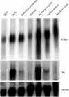
Figure 1
Figure 1
Northern blot analysis of KLK8 mRNA in normal skin and the lesional skin of patients with cutaneous diseases. The same blot was rehybridised with the tissue plasminogen activator (tPA) probe and with a glyceralaldehyde 3-phosphate dehydrogenase (GAPDH) probe. Note the different band patterns between KLK8 and tPA mRNAs in the various skin samples. BCC, basal cell carcinoma; SCC, squamous cell carcinoma.
Figure 2
Figure 2
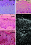
In situ hybridisation for KLK8 mRNA in normal and pathological skin samples. (A) In normal skin, weak hybridisation signals were localised only in the superficial layer shown by arrows. No signals and strong hybridisation signals were found in the lesional skin samples of psoriasis vulgaris using (B) sense and (C) antisense probes for KLK8 mRNA, respectively. Parakeratotic cells localised in the cornified layer did not express KLK8 mRNA (arrowheads). (E) Strong hybridisation signals (asterisk) for KLK8 mRNA were found in lesional skin of seborrheic keratosis. (A, B, C, And E) are bright field micrographs of skin sections counterstained by haematoxylin and eosin; (D) and (F) are dark field micrographs of the same fields shown in (C) and (E), respectively. Scale bars: (A), 50 μm; (B–F), 100 μm.
Figure 3
Figure 3
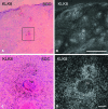
In situ hybridisation for KLK8 mRNA in pathological skin. In situ hybridisation of KLK8 mRNA in the lesional skin of (A and B) lichen planus and (C and D) marginal skin around a decubitus ulcer. (A) And (C) are bright field micrographs of skin sections counterstained with haematoxylin and eosin; (B) and (D) are dark field micrographs of the same fields shown in (A) and (B), respectively. Silver grains were observed under light and dark field illumination (A–D). Small arrows in (A) and (B) represent cells labelled by the hybridisation probe. Thick arrows (yellow) represent artifacts (D). Scale bars: (A,B), 100 μm; (C,D), 200 μm.
Figure 4
Figure 4
In situ hybridisation of KLK8 mRNA in the lesional skin of squamous cell carcinoma (SCC). (A) Low power micrograph showing haematoxylin and eosin staining of highly differentiated SCC. (B) Dark field micrograph showing silver grains around horn pearls (asterisks). (C) And (D) are high power magnifications of the boxed area in (A) showing intense signals for KLK8 mRNA. (A) And (C) are bright field micrographs of skin sections counterstained by haematoxylin and eosin; (B) and (D) are dark field micrographs of the same fields shown in (A) and (C), respectively. Scale bars: (A,B), 1 mm; (C,D), 400 μm. hp, horn pearl.
Figure 5
Figure 5
Northern blot analysis of KLK8 and tissue type plasminogen activator (tPA) mRNA in cultured human keratinocytes. Total RNA was collected at the stage of 70% confluence (70%) and 100% confluence (100%) in low Ca2+ medium (low Ca2+). Total RNA was also collected at one day (1D) and three days (3D) of culture after changing to high Ca2+ medium (high Ca2+). KLK8 mRNA expression increased greatly in parallel with the differentiation of keratinocytes. GAPDH, glyceralaldehyde 3-phosphate dehydrogenase.

Similar articles

Cited by

References

    1. Zelickson AS. Ultrastructure of normal and abnormal skin. Philadelphia: Lea & Febiger, 1967.
    1. Inoue N, Kuwae K, Ishida-Yamamoto A, et al. Expression of neuropsin in the keratinizing epithelial tissue—immunohistochemical analysis of wild-type and nude mice. J Invest Dermatol 1998;110:923–31. - PubMed
    1. Jensen PJ, Lavker RM. Urokinase is a positive regulator of epidermal proliferation in vivo. J Invest Dermatol 1999;112:240–4. - PubMed
    1. Weil M, Raff MC, Braga VM. Caspase activation in the terminal differentiation of human epidermal keratinocytes. Curr Biol 1999;9:361–4. - PubMed
    1. Isseroff RR, Fusenig NE, Rifkin DB. Plasminogen activator in differentiating mouse keratinocytes. J Invest Dermatol 1983;80:217–22. - PubMed

Publication types

MeSH terms