. 2002 Aug;55(4):250-61.
doi: 10.1136/mp.55.4.250.

A role for CCN3 (NOV) in calcium signalling

Affiliations

A role for CCN3 (NOV) in calcium signalling

C L Li et al. Mol Pathol. 2002 Aug.

Abstract

Aims: In animals and humans increased expression of CCN3 (NOV) is detected in tissues where calcium is a key regulator, such as the adrenal gland, central nervous system, bone and cartilage, heart muscle, and kidney. Because the multimodular structure of the CCN proteins strongly suggests that these cell growth regulators are metalloproteins, this study investigated the possible role of CCN3 in ion flux and transport during development, control of cell proliferation, differentiation, and pathobiology.

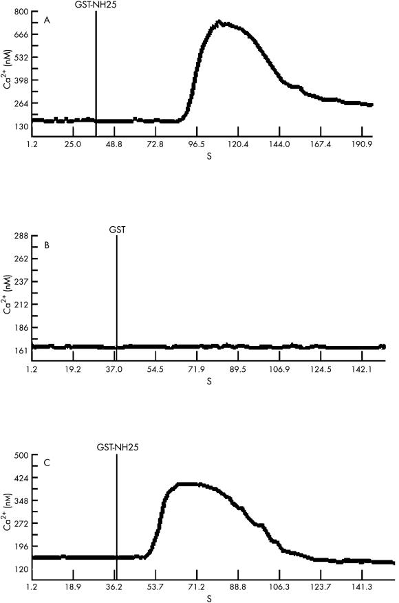
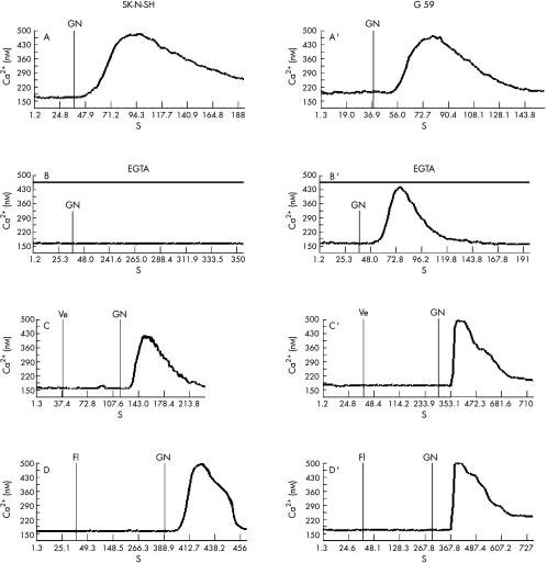
Methods: The isolation of CCN3 partners was performed by means of the two hybrid system. Yeasts were cotransfected with an HL60 cDNA library fused to the transactivation domain of the GAL4 transcription factor, and with a plasmid expressing CCN3 fused to the DNA binding domain of GAL4. Screening of the recombinant clones selected on the basis of leucine, histidine, and tryptophan prototrophy was performed with a beta-galactosidase assay. After the interaction between CCN3 and its putative partners was checked with a GST (glutathione S-transferase) pull down assay, the positive clones were identified by cloning. To establish whether the CCN3 protein affected calcium ion flux, a dynamic imaging microscopy system was used, which allowed the fluorometric measurement of the intracellular calcium concentration. The proteins used in the assays were GST fused with either CCN3 or CCN2 (CTGF) and GST alone as a control.

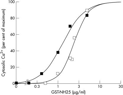
Results: The two hybrid system identified the S100A4 (mts1) calcium binding protein as a partner of CCN3 and the use of the GST fusion proteins showed that the addition of CCN3 and CCN2 to G59 glioblastoma and SK-N-SH neuroblastoma cells caused a pronounced but transient increase of intracellular calcium, originating from both the entry of extracellular calcium and the mobilisation of intracellular stores.

Conclusions: The interaction of CCN3 with S100A4 may account, in part, for the association of CCN3 with carcinogenesis and its pattern of expression in normal conditions. The increased intracellular calcium concentrations induced by CCN3 and CCN2 both involve different processes, among which voltage independent calcium channels might be of considerable importance in regulating the calcium flux associated with cell growth control, motility, and spreading. These observations assign for the first time a biological function to the CCN3 protein and point out a broader role for the CCN proteins in calcium ion signalling.


Figures

Figure 1
Figure 1
Part 1: Amino acid sequence alignment in CCN3 proteins. The amino acid sequence of CCN3 proteins from different species has been aligned with the interalign program (University of Sandford, USA). Those residues whose position is conserved throughout evolution are shaded in black. The 38 conserved cysteine residues and conserved prolines are shaded in grey. The position and sequences of the TSP1 (thrombospondin type 1 repeat) and CT (cystin knot) motifs have been indicated in bold above the CCN3 consensus sequences for each domain.
Figure 1
Figure 1
Part 2: Amino acid sequence alignment in CCN3 proteins. The amino acid sequence of CCN3 proteins from different species has been aligned with the interalign program (University of Sandford, USA). Those residues whose position is conserved throughout evolution are shaded in black. The 38 conserved cysteine residues and conserved prolines are shaded in grey. The position and sequences of the IB (insulin-like growth factor binding related proteins) and VWC (von willebrand type C repeat) motifs have been indicated in bold above the CCN3 consensus sequences for each domain.
Figure 2
Figure 2
Distribution of sequences encoding the ccn3 gene and multimodular organisation of the CCN3 protein. (A) Schematic representation of the exon distribution in the human ccn3 gene. The sizes are given in base pairs. Boxed sequences are present in the human ccn3 cDNA clone used in previous studies (see Perbal for review). (B) Schematic representation of the five structural modules of the CCN3 protein. The signal peptide (SP) is encoded by exon 1 (e1) and the domains 1–4 (D1–4) are encoded by exons 2–5 (e2–5). The length of each domain is indicated in amino acid residues. The number of cysteine residues present in each domain is shown in square brackets. The positions of amino acid sequence motifs sharing identity with insulin-like growth factor binding related proteins (IB), von Willebrand type C repeat (VWC), thrombospondin type 1 repeat (TSP1), and cystin knot (CT) are indicated below each module. The consensus sequence for each motif is indicated in fig 1 ▶ ▶.
Figure 3
Figure 3
Organisation of the S100A4 sequences in clone CLM. (A) The nucleotide sequence of the GAL4AD-CLM clone at the cloning site and (B) the protein encoded by the GAL4AD-CLM clone at the cloning site. The pGADGH vector sequences are indicated in italics. The HeLa cDNA transcripts were cloned at the EcoRI site. The nucleotide sequence contained between e1b and e2 corresponds to the alternative 5` non-coding sequences characterised in human cells. The S100A4 coding sequences are contained in exons 2 and 3. The two sets of sequences in bold correspond to the calcium binding loops in the S100A4 protein. The first calcium binding site, which is encoded by exon 2, is composed of 14 amino acids with a helix–loop–helix conformation (helix A: VMVSTFHKY; loop 1: SGKEGDKFKLNKSE; helix B: LKELLTR). It corresponds to the binding loop of the pseudo EF hand (from V at position 11 to E at position 41) characteristic of the S100 proteins. The second calcium binding site, encoded by exon 3, is composed of 12 amino acids (helix C: AFQKLMSNL; loop 2: DSNRDNEVDFQE; helix D: YCVFLSC) contained in a canonical EF hand (from F at position 52 to I at position 82).
Figure 4
Figure 4
Effect of GST–NH25 on intracellular calcium increase in G59 and SK-N-SH cells. (A) The GST–NH25 protein (1 μg/ml) was added to G59 cells after 40 seconds of background recording. (B) GST only (5 μg/ml) was tested as control on G59 cells. (C) Under the same conditions, GST–NH25 (2 μg/ml) was applied to SK-N-SH cells.
Figure 5
Figure 5
Intracellular calcium mobilisation induced by increasing concentrations of GST–NH25. Neuroblastoma SK-N-SH (open squares) or glioblastoma G59 (closed squares) cells were treated with increasing concentrations of GST–NH25 protein. The intracellular calcium influx was determined for each concentration, and represented as the percentage of the maximal activation. All other details are as described under the materials and methods section.
Figure 6
Figure 6
Effects of calcium blockers on intracellular calcium mobilisation induced by GST–NH25 in (A–D) SK-N-SH and (A’–D’) G59 cells. GST–NH25 (2 μg/ml) was applied in (A,A’) the absence or the presence of (B,B’) 20mM EGTA, (C,C’) 1μM verapamil, or (D,D’) 10μM flunarizine.

Similar articles

Cited by

References

    1. Bork P. The modular architecture of a new family of growth regulators related to connective tissue growth factor. FEBS Lett 1993;327:125–30. - PubMed
    1. Lau LF, Lam SC. The CCN family of angiogenic regulators: the connection. Exp Cell Res 1999;248:44–57. - PubMed
    1. Brigstock DR. The connective tissue growth factor/cysteine-rich 61/nephroblastoma overexpressed (CCN) family. Endocr Rev 1999;20:189–206. - PubMed
    1. Perbal B. NOV (nephroblastoma overexpressed) and the CCN family of genes: structural and functional issues. Mol Pathol 2001;54:57–79. - PMC - PubMed
    1. Hashimoto BY, Shindo-Okada N, Tani M, et al. Expression of the ELM1 gene, a novel gene of the CCN (connective tissue growth factor, Cyr61/Cef10, and neuroblastoma overexpressed gene) family, suppresses in vivo tumor growth and metastasis of K-1735 murine melanoma cells. J Exp Med 1998;187:289–96. - PMC - PubMed

Publication types

MeSH terms