. 2002 Aug;55(4):262-72.
doi: 10.1136/mp.55.4.262.

Alterations of the MDV oncogenic regions in an MDV transformed lymphoblastoid cell line

Affiliations

Alterations of the MDV oncogenic regions in an MDV transformed lymphoblastoid cell line

E Le Rouzic et al. Mol Pathol. 2002 Aug.

Abstract

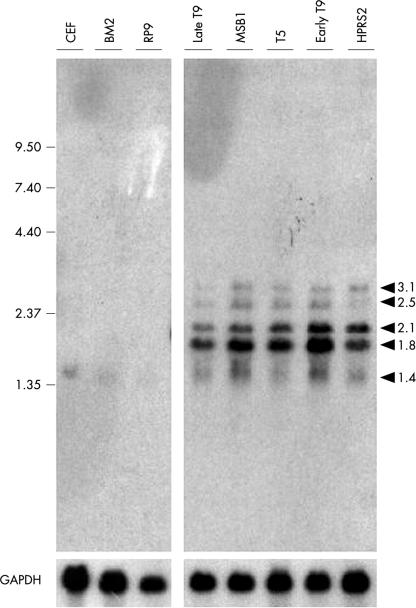
Aims: Lymphoblastoid cell lines derived from Marek's disease virus (MDV) induced tumours have served as models of MDV latency and transformation. They are stable and can be cultured with no detectable MDV genomic alterations upon repeated passaging. An MDV transformed lymphoblastoid T cell line (T9 cell line) has been reported to contain a disrupted MDV BamHI-H fragment and a Rous associated virus insertional activation of the c-myb protooncogene. In an attempt to define the respective participation of c-myb and MDV in the transformed phenotype of T9 cells, an analysis of MDV oncogenic sequences (BamHI-H, BamHI-A, and EcoQ fragments) was performed in these cells.

Methods: Using two different passages of the T9 cell line (late and early passages), the organisation of the MDV oncogenic regions and their expression in these cells were analysed. In vivo assessment of the oncogenicity of the virus contained within these cells was assessed by injecting them into 1 day old chickens.

Results: In T9 cells maintained in culture for up to six months (late T9), the MDV ICP4 gene was disrupted, whereas the meq gene was actively transcribed. The alterations of the MDV genome in these cells correlated with the inability of the virus to induce the classic signs of Marek's disease in 1 day old chickens. However, early T9 cells submitted to a limited number of passages induced classic MDV pathogenicity, as efficiently as the MDV control cell line (T5), and did not show gross structural changes in the oncogenic MDV sequences.

Conclusions: Although the expression pattern of the MDV oncogenes in early T9 cells was identical to the one reported for other MDV transformed cells, longterm culture of an MDV transformed cell line containing a RAV insertional activation of the c-myb protooncogene led to the disruption of the MDV BamHI-H and BamHI-A oncogenic regions. In the late T9 cells MEQ was the only detected MDV oncoprotein. These results suggest that in the late T9 cells the truncated MYB protein compensates for the loss of MDV oncoproteins and reinforce the possibility that MEQ and MYB cooperate in the maintenance of the transformed state and the tumorigenic potential of these cells.


Figures

Figure 1
Figure 1
(A) Genomic structure of Marek’s disease virus (MDV). MDV consists of long and short unique sequences (UL and US, respectively), flanked by long and short internal repeats (IRL and IRS, respectively), and long and short inverted terminal repeats (TRL and TRS, respectively). (B) Locations of the BamHI-D, BamHI-H, BamHI-I2, BamHI-Q2, BamHI-L, and BamHI-A regions. A detailed restriction map is indicated (E, EcoRI; B, BamHI; P, PvuII). (C) Locations of the pp24, pp38, meq, and ICP4 homologue genes. Corresponding gene regions are enlarged to show the open reading frame and the transcription sense (arrow); the localisations of the oligonucleotides (arrowhead) used for PCR and Southern blotting are indicated. A solid bar represents the resulting amplified product used as a probe.
Figure 2
Figure 2
Disruption of Marek’s disease virus (MDV) BamHI-H fragment in late T9 DNA. Southern blots of BamHI digested high molecular weight DNA (15 μg) were hybridised with the [32P] labelled MDV specific 5.4 kb BamHI-H probe. DNA samples from chicken embryo fibroblasts (CEFs) were used as negative control. Molecular weight markers (in kilobases) are from HindIII digested λ DNA.
Figure 3
Figure 3
Analysis of the ICP4 gene region. Expression of RNA species in Marek’s disease virus (MDV) transformed cell lines. Northern blots were performed with 20 μg samples of total RNA, and were sequentially probed with [32P] labelled ICP4 and glyceraldehyde-3-phosphate dehydrogenase (GAPDH) double strand fragments. Chicken embryo fibroblasts (CEFs) and BM2 RNA samples were used as negative controls. Size markers in kilobases correspond to the BRL RNA ladder.
Figure 4
Figure 4
Analysis of the ICP4 homologue gene region. Southern blot analysis. DNA (15 μg) digested with EcoRI (left hand panel) or PvuII (right hand panel) from the indicated cells was run on a 0.8% agarose gel and then blotted and hybridised with the ICP4 probe. Molecular weight markers (in kilobases) are from HindIII digested λ DNA. CEF, chicken embryo fibroblast.
Figure 5
Figure 5
Analysis of the Eco-Q region encoding the meq gene. Southern blot analysis. DNA (15 μg) digested with BamHI (left hand panel) or EcoRI (right hand panel) from the indicated cells was blotted and hybridised with the meq probes. Chicken embryo fibroblast (CEF) and RP9 digested DNA samples were used as negative controls. Molecular weight markers (in kilobases) are from HindIII digested λ DNA.
Figure 6
Figure 6
Analysis of the Eco-Q region encoding the meq gene. The expression of meq RNA species in Marek’s disease virus transformed cell lines. Northern blots were performed with 20 μg samples of total RNA from the indicated cells, and were sequentially hybridised with [32P] labelled probes meq and glyceraldehyde-3-phosphate dehydrogenase (GAPDH) double strand fragments. Chicken embryo fibroblast (CEF) and BM2 RNA samples were used as negative controls. The size markers (in kilobases) correspond to the BRL RNA ladder.
Figure 7
Figure 7
Analysis of the Eco-Q region encoding the meq gene. The expression of MEQ proteins in Marek’s disease virus (MDV) transformed cell lines. Proteins (100 μg) were denatured in Laemmli buffer and then resolved on 10% sodium dodecyl sulfate-polyacrylamide gel electrophoresis before transferring on to a PVDF membrane. The blot was probed with rabbit anti-MEQ polyclonal antibodies (1/3000 dilution27), followed by detection using the enhanced chemiluminescence (ECL) system (Amersham). Cell extracts from chicken embryo fibroblasts (CEFs) and BM2 were used as negative controls. Specific MDV products are indicated by arrows. Prestained molecular weight markers (Novex) are indicated in kilodaltons.
Figure 8
Figure 8
Molecular analysis of the tumours (or normal tissues) isolated from T5, early T9, and late T9 cell line inoculated and contact chickens. (A) Detection of Marek’s disease virus (MDV) sequences in DNA from induced tumours. PCR amplification was carried out on DNA isolated from different tissues of chickens injected with T5 (964, 955, and 959), early T9 (479 and 480), and late T9 (971 and 980). The control without DNA template (lane C) was performed under identical conditions. Additional controls included DNA isolated from the early T9, late T9, and T5 cell lines. PCR products were run on a 1.5% agarose gel and visualised with an ultraviolet transilluminator. The positions of selected bands from a 1 kb ladder marker (BRL) are indicated on the left. (B) PCR products run on the gel were transferred on to a nitrocellulose membrane and then hybridised with [32P] labelled oligonucleotides pp38I. (C) PCR amplification of the 5` RAV–c-myb junction. The same DNA templates as in (A) were subjected to 30 rounds of amplification using primer 1 (corresponding to c-myb exon 3 sequences) and primer 2 (corresponding to RAV gag sequences). PCR products were run on a 1.5% agarose gel and visualised by means of an ultraviolet transilluminator. The position of the PCR amplified 850 bp 5` junction fragment is indicated. Control experiments were performed under identical conditions without DNA (lane C).

Similar articles

References

    1. Biggs PM. The Leeuwenhoek Lecture. Marek’s disease herpesvirus: oncogenesis and prevention. Philos Trans R Soc Lond B Biol Sci 1997;352:1951–62. - PMC - PubMed
    1. Fukuchi K, Sudo M, Lee Y, et al. Structure of Marek’s disease virus DNA: detailed restriction enzyme map. J Virol 1984;51:102–9. - PMC - PubMed
    1. Tulman ER, Afonso CL, Lu Z, et al. The genome of a very virulent Marek’s disease virus. J Virol 2000;74:7980–8. - PMC - PubMed
    1. Lee LF, Wu P, Sui D, et al. The complete unique long sequence and the overall genomic organization of the GA strain of Marek’s disease virus. Proc Natl Acad Sci U S A 2000;97:6091–6. - PMC - PubMed
    1. Ross NL. T-cell transformation by Marek’s disease virus. Trends Microbiol 1999;7:22–9. - PubMed

Publication types