

Case Reports
doi: 10.1128/JCM.40.8.3071-3075.2002.
Corneal ulcer caused by the new fungal species Sarcopodium oculorum
Affiliations
- PMID: 12149384
- PMCID: PMC120633
- DOI: 10.1128/JCM.40.8.3071-3075.2002
Item in Clipboard
Case Reports
Corneal ulcer caused by the new fungal species Sarcopodium oculorum
J Clin Microbiol.
2002 Aug.
Abstract
We describe a case of keratitis caused by a new species of the hyphomycetous genus Sarcopodium, S. oculorum. The corneal ulcer developed after 5 months of treatment with corticosteroids in a Brazilian boy diagnosed with allergic conjunctivitis. Fungal hyphae and conidia were detected in corneal scrapings, and repeated cultures were positive for this fungus. The infection was resolved with natamycin and ketoconazole. Eleven antifungals were tested against this fungus, and all except flucytosine and fluconazole showed in vitro activity.
Figures
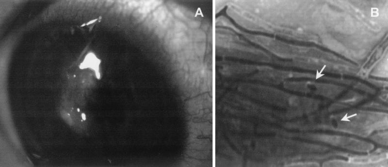
(A) Corneal ulcer. (B) Gram stain showing segmented hyphae and conidia (arrows). Magnification, ×1,280.
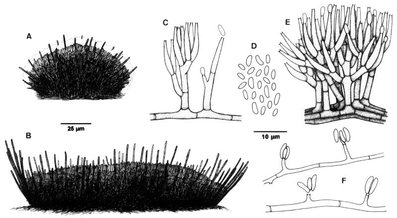
S. oculorum IMI 387421. (A and B) Sporodochia. (C to E) Conidiophores and conidia from sporodochia. (F) Undifferentiated hyphae with conidiogenous cells and conidia.
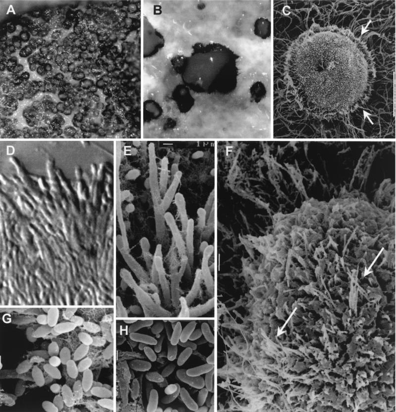
S. oculorum IMI 387421. (A and B) Sporodochia from the colony growing on PDA after 3 weeks of incubation at 25°C. (C) Sporodochium with setae (arrows). (D and E) Conidiophores from sporodochia. (F) Part of a sporodochium showing setae (arrows) among conidiophores. (G) Conidia from sporodochia. (H) Conidia from undifferentiated hyphae. Magnifications: ×120 (A), ×400 (B), ×200 (C), ×2,130 (D), ×3,010 (E), ×600 (F), and ×4,000 (G and H).
Similar articles
-
Fungal keratitis caused by Scopulariopsis brevicaulis treated successfully with natamycin.Cornea. 2004 Mar;23(2):201-3. doi: 10.1097/00003226-200403000-00015. Cornea. 2004. PMID: 15075891
-
Medical management of Beauveria bassiana keratitis.Cornea. 2000 May;19(3):405-6. doi: 10.1097/00003226-200005000-00031. Cornea. 2000. PMID: 10832710
-
Series of five cases of Papulaspora equi keratomycosis.Cornea. 2014 Jun;33(6):640-3. doi: 10.1097/ICO.0000000000000108. Cornea. 2014. PMID: 24699559
-
Corneal ulcer due to a rare pleosporalean member of the genus Bipolaris following cow tail injury to the eye: A case report and review of literature.Indian J Ophthalmol. 2017 May;65(5):403-405. doi: 10.4103/ijo.IJO_836_16. Indian J Ophthalmol. 2017. PMID: 28573998 Free PMC article. Review.
-
Paecilomyces lilacinus keratitis: two case reports in extended wear contact lens wearers.CLAO J. 1987 Mar-Apr;13(2):95-101. CLAO J. 1987. PMID: 3329072 Review. No abstract available.
Cited by
-
Keratitis due to the wood saprobic ascomycete, Auerswaldia lignicola (Family Botryosphaeriaceae), in a carpenter in India.Mycopathologia. 2013 Dec;176(5-6):463-6. doi: 10.1007/s11046-013-9713-5. Epub 2013 Oct 26. Mycopathologia. 2013. PMID: 24158617
-
Subcutaneous phaeohyphomycosis due to Phialemoniopsis ocularis successfully treated by voriconazole.Med Mycol Case Rep. 2014 May 10;5:4-8. doi: 10.1016/j.mmcr.2014.04.001. eCollection 2014 Jul. Med Mycol Case Rep. 2014. PMID: 24936402 Free PMC article.
-
New pyrenochaeta species causing keratitis.J Clin Microbiol. 2009 May;47(5):1596-8. doi: 10.1128/JCM.01912-08. Epub 2009 Mar 18. J Clin Microbiol. 2009. PMID: 19297598 Free PMC article.
-
Case of keratitis caused by an uncommon Fusarium species.J Clin Microbiol. 2003 Dec;41(12):5823-6. doi: 10.1128/JCM.41.12.5823-5826.2003. J Clin Microbiol. 2003. PMID: 14662993 Free PMC article.
-
Subcutaneous phaeohyphomycotic nodule due to Phialemoniopsis hongkongensis sp. nov.J Clin Microbiol. 2014 Sep;52(9):3280-9. doi: 10.1128/JCM.01592-14. Epub 2014 Jun 25. J Clin Microbiol. 2014. PMID: 24966363 Free PMC article.
References
-
- Behrens-Braumann, W. 1999. Mycosis of the eye and its adnexa. Dev. Ophthalmol. 32:i-ix, 1-201. - PubMed
-
- De Hoog, G. S., J. Guarro, J. Gené, and M. J. Figueras. 2000. Atlas of clinical fungi, 2nd ed. Centraalbureau voor Schimmelcultures, Utrecht, The Netherlands, and the Rovira i Virgili University, Reus, Spain.
-
- Ellis, M. B. 1976. More dematiaceous hyphomycetes. Commonwealth Mycological Institute, Kew, United Kingdom.
-
- Foster, C. S. 1992. Fungal keratitis. Infect. Dis. Clin. N. Am. 6:851-857. - PubMed
-
- Gené, J., and J. Guarro. 1996. A new Chaetomium from Thailand. Mycol. Res. 100:1005-1009.
Publication types
MeSH terms
Substances
LinkOut - more resources
Full Text Sources
Medical
