. 2002 Aug 6;99(16):10825-30.
doi: 10.1073/pnas.152112399. Epub 2002 Jul 29.

Reduced fear expression after lesions of the ventral hippocampus

Affiliations

Reduced fear expression after lesions of the ventral hippocampus

Kirsten G Kjelstrup et al. Proc Natl Acad Sci U S A. .

Abstract

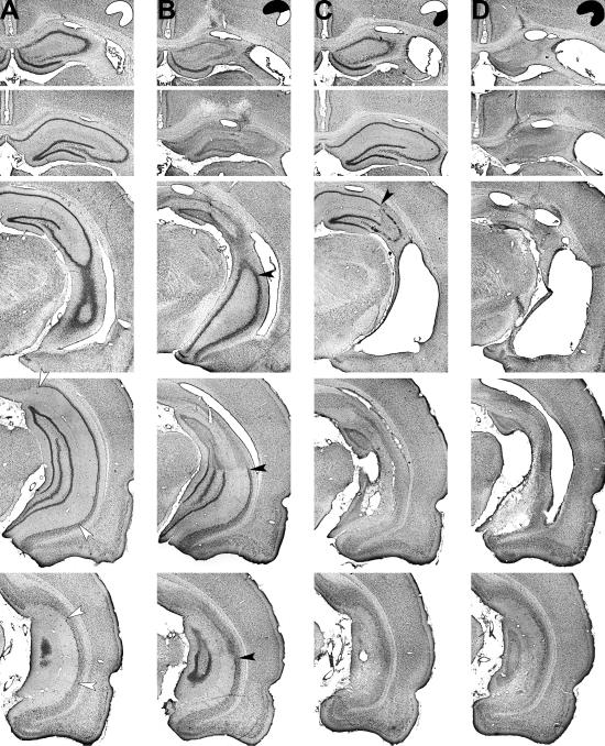
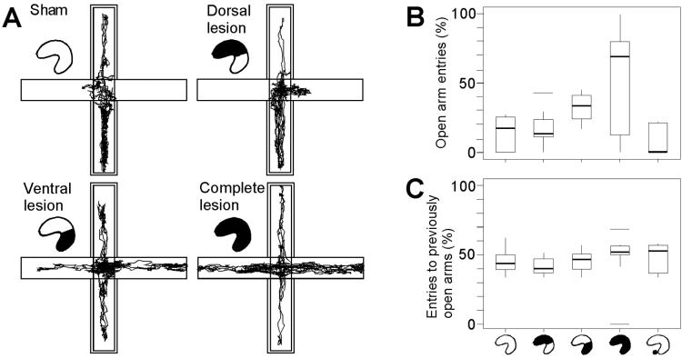
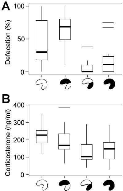
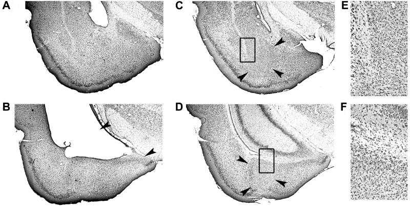
The hippocampus has a critical role in several fundamental memory operations, including the conditioning of fear to contextual information. We show that the hippocampus is necessary also for unconditioned fear, and that the involved circuitry is at the ventral pole of the hippocampus. Rats with selective hippocampal lesions failed to avoid open arms in an elevated plus-maze and had decreased neuroendocrine stress responses during confinement to a brightly lit chamber. These effects were reproduced by lesions of the ventral half of the hippocampus, but not by damage to the dorsal three-quarters of the hippocampus or the amygdala. Ventral lesions failed to impair contextual fear conditioning or spatial navigation, suggesting that the ventral hippocampus may specifically influence some types of defensive fear-related behavior.


Figures

Fig 1.
Fig 1.
Representative cresyl violet stains of intact neuronal cell bodies at five coronal levels through the hippocampus after sham surgery (A) or ibotenate lesions of the dorsal hippocampus (B), ventral hippocampus (C), or entire hippocampus (D). Filled arrowheads, borders between lesioned and healthy tissue; open arrowheads, borders of the subiculum.
Fig 2.
Fig 2.
Reduced open-arm avoidance after selective lesions of the ventral hippocampus. (A) Paths of representative animals with sham operations or dorsal, ventral, or complete lesions of the hippocampus, during 10 min of exposure to an elevated plus maze with two open arms. Double contours indicate enclosed arms. (B) Box plot comparing performance in rats with dorsal, ventral, or complete hippocampal lesions, or lesions of the adjacent parts of the amygdala (hippocampal icons as in Fig. 1; amygdala group to the right). The diagram shows median percentage of visits to open arms (thick horizontal line inside box), interquartile distances (boxes), upper and lower limits [Q1 − 1.5 (Q3–Q1) and Q3 + 1.5 (Q3–Q1), where Q1 and Q3 are first and third quartiles, respectively], and outliers (horizontal lines). (C) Visits to previously open arms on a subsequent trial with all arms enclosed.
Fig 3.
Fig 3.
Selective lesions of the ventral hippocampus reduced defecation and corticosterone secretion during exposure to a brightly lit test chamber (box plots; symbols as in Fig. 2). (A) Percentage of trials during which rats left one or several feces during confinement to a brightly lit test chamber. (B) Plasma corticosterone concentration 20 min after exposure to the test chamber.
Fig 4.
Fig 4.
Coronal sections showing representative cresyl violet stains including amygdala and ventral hippocampus after ibotenate lesions targeted either at the ventral hippocampus (A and B) or the adjacent nuclei of the amygdala (CF). Arrowheads indicate approximate borders between lesioned and intact tissue. Transitions between intact and lesioned tissue in C and D (boxes) are shown at high magnification in E and F, respectively. The behavior of rats with lesions in the amygdala is shown in Fig. 2.
Fig 5.
Fig 5.
Intact contextual and spatial memory after lesions of the ventral hippocampus. (A) Percentage of freezing (means ± SEM) during the first 5 min of confinement to a test chamber before and 24 h after the rats received electric foot shock in this chamber. (B) Retention on a probe trial at the beginning of day 3 in the water maze. The diagram shows the time that rats spent in a circular zone around the platform position (black) and in corresponding zones of the three other quadrants (means ± SEM). Each zone covered 4.1% of the pool surface (radius, 20 cm; expected time, 2.5 s; Inset).

References

    1. Davis M., Walker, D. L. & Lee, Y. (1997) Philos. Trans. R. Soc. London B 352, 1675-1687. - PMC - PubMed
    1. Fendt M. & Fanselow, M. S. (1999) Neurosci. Biobehav. Rev. 23, 743-760. - PubMed
    1. LeDoux J. E. (2000) Annu. Rev. Neurosci. 23, 155-184. - PubMed
    1. Maren S. (2001) Annu. Rev. Neurosci. 24, 897-931. - PubMed
    1. Aggleton J. P., (2000) The Amygdala: A Functional Analysis (Oxford Univ. Press, Oxford).

Publication types

MeSH terms