. 2002 Aug 6;99(16):10260-5.
doi: 10.1073/pnas.152346599. Epub 2002 Jul 29.

De novo determination of peptide structure with solid-state magic-angle spinning NMR spectroscopy

Affiliations

De novo determination of peptide structure with solid-state magic-angle spinning NMR spectroscopy

Chad M Rienstra et al. Proc Natl Acad Sci U S A. .

Abstract

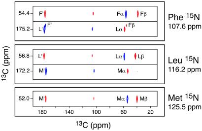
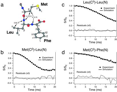
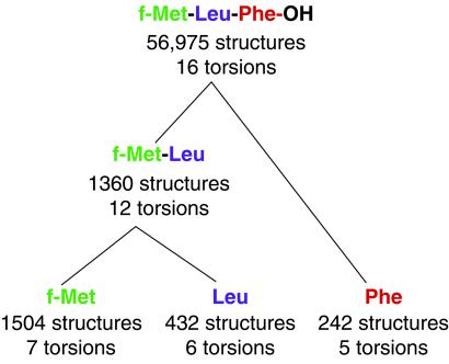
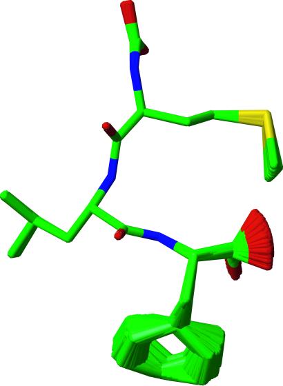
The three-dimensional structure of the chemotactic peptide N-formyl-l-Met-l-Leu-l-Phe-OH was determined by using solid-state NMR (SSNMR). The set of SSNMR data consisted of 16 (13)C-(15)N distances and 18 torsion angle constraints (on 10 angles), recorded from uniformly (13)C,(15)N- and (15)N-labeled samples. The peptide's structure was calculated by means of simulated annealing and a newly developed protocol that ensures that all of conformational space, consistent with the structural constraints, is searched completely. The result is a high-quality structure of a molecule that has thus far not been amenable to single-crystal diffraction studies. The extensions of the SSNMR techniques and computational methods to larger systems appear promising.


Figures

Fig 1.
Fig 1.
Strip cross sections through the 15N planes of the 3D 15N–13C–13C chemical shift correlation spectrum of f-MLF-OH, showing the backbone resonance assignments. The Met 15N plane (125.5 ppm) shows only 13C cross-peaks from the Met residue. In contrast, the Leu 15N plane (116.2 ppm) shows Met and Leu 13C cross-peaks, and the Phe 15N plane (107.6 ppm) displays Leu and Phe 13C cross-peaks. Since 13C–13C correlations were established by using the SPC-5 double-quantum recoupling pulse sequence (31) the cross-peaks corresponding to subsequent 13C–13C dipolar transfers alternate in sign (28) (blue and red for positive and negative absorption, respectively). Details of the pulse sequence and experimental parameters used to record this spectrum can be found in ref. .
Fig 2.
Fig 2.
Measurement of ψMet in f-MLF-OH by the double-quantum 15N–13C–13C–15N experiment (38). (a) Experimental and simulated dephasing of the Met C′–Cα double-quantum coherence under the C′–N and Cα–N dipolar couplings. The best simulation yields a torsion angle of ±157° ± 1°. (b) rmsd between the NCCN simulation and experiment for the Met residue, calculated as a function of ψ.
Fig 3.
Fig 3.
Measurement of carbon–nitrogen internuclear distances in [U-13C,15N]f-MLF-OH by frequency-selective REDOR (16). (a) Structural model of f-MLF-OH displaying the distances measured in b–d. Experimental REDOR S/S0 curves (S0 and S represent the reference and dipolar dephasing experiments, respectively) and simulations are shown for Met(Cβ)–Leu(N) (b), Leu(Cδ)–Leu(N) (c), and Met(Cβ)–Phe(N) (d), and they correspond to internuclear distances of 3.12 ± 0.03 Å (b), 3.64 ± 0.09 Å (c), and 4.12 ± 0.15 Å (d). A total of 16 distances between 2.5 and 6 Å were measured in f-MLF-OH. Distance measurements were performed in a sample prepared by cocrystallizing [U-13C,15N]f-MLF-OH with natural-abundance f-MLF-OH in a 1:9 ratio, to minimize the interference from intermolecular 13C–15N couplings. Details of the pulse sequence and experimental parameters can be found in ref. .
Fig 4.
Fig 4.
An illustration of the divide-and-conquer strategy used to search conformational space. Starting from the individual residues (bottom) and progressing to the tripeptide, the number of substructures satisfying the SSNMR and excluded-volume constraints and the number of searchable torsions are indicated.
Fig 5.
Fig 5.
An illustration of a family of nearly identical structures that were filtered from the total of 56,975 f-MLF-OH structures for ease of display. The set shown is representative of the entire ensemble and is consistent with the SSNMR torsion angle measurements, 13C–15N distances, and excluded-volume constraints. The structure of the backbone is of especially high quality (0.02 Å rmsd). Since the formyl group was not labeled, it was permitted to assume both the cis and trans conformations in the calculation, and it exhibits the appearance of a carboxyl group in the figure. The carboxyl terminus and the Phe ring appear disordered because no torsion angle methods currently exist to constrain the terminal ψ or χ2 angle. The ring conformation is largely determined by excluded volume constraints, and it is likely undergoing twofold flips (see text). The Met and Leu side-chain conformations are also relatively well defined.

Similar articles

Cited by

References

    1. Wüthrich K., (1986) NMR of Proteins and Nucleic Acids (Wiley, New York).
    1. Gayathri C., Bothner-By, A. A., van Zijl, P. C. M. & MacLean, C. (1982) Chem. Phys. Lett. 87, 192-196.
    1. Tolman J. R., Flanagan, J. M., Kennedy, M. A. & Prestegard, J. H. (1995) Proc. Natl. Acad. Sci. USA 92, 9279-9283. - PMC - PubMed
    1. Tjandra N. & Bax, A. (1997) Science 278, 1111-1114. - PubMed
    1. Pervushin K., Riek, R., Wider, G. & Wüthrich, K. (1997) Proc. Natl. Acad. Sci. USA 94, 12366-12371. - PMC - PubMed

Publication types

MeSH terms

Substances

LinkOut - more resources