. 2002 Aug 6;99(16):10718-23.
doi: 10.1073/pnas.152597199. Epub 2002 Jul 29.

Up-regulation of plasma membrane-associated ganglioside sialidase (Neu3) in human colon cancer and its involvement in apoptosis suppression

Affiliations

Up-regulation of plasma membrane-associated ganglioside sialidase (Neu3) in human colon cancer and its involvement in apoptosis suppression

Yoichiro Kakugawa et al. Proc Natl Acad Sci U S A. .

Abstract

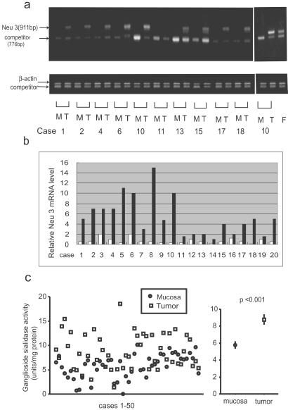
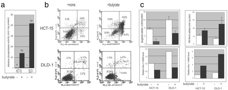
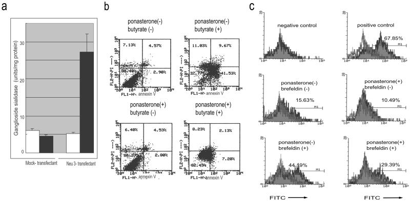
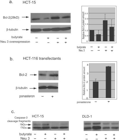
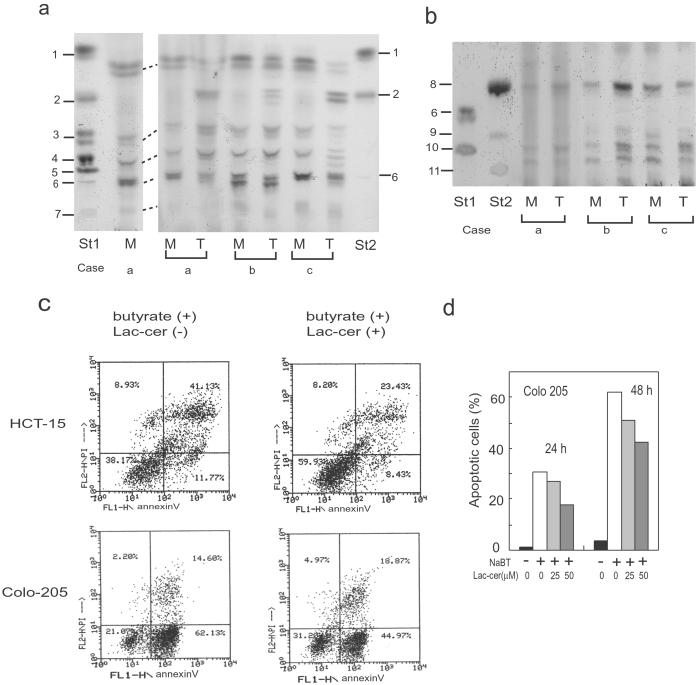
Human plasma membrane-associated sialidase (Neu3) is unique in specifically hydrolyzing gangliosides, thought to participate in cell differentiation and transmembrane signaling, thereby playing crucial roles in the regulation of cell surface functions. We have discovered levels of mRNA for this sialidase to be increased in restricted cases of human colon cancer by 3- to 100-fold compared with adjacent nontumor mucosa (n = 32), associated with significant elevation in sialidase activity in tumors (n = 50). In situ hybridization showed the sialidase expression in epithelial elements of adenocarcinomas. In cultured human colon cancer cells, the sialidase level was down-regulated in the process of differentiation and apoptosis induced by sodium butyrate, whereas lysosomal sialidase (Neu1) was up-regulated. Transfection of the sialidase gene into colon cancer cells inhibited apoptosis and was accompanied by increased Bcl-2 and decreased caspase expression. Colon cancer exhibited a marked accumulation of lactosylceramide, a possible sialidase product, and addition of the glycolipid to the culture reduced apoptotic cells during sodium butyrate treatment. These results indicate that high expression of the sialidase in cancer cells leads to protection against programmed cell death, probably modulation of gangliosides. This finding provides a possible sialidase target for diagnosis and therapy of colon cancer.


Figures

Fig 1.
Fig 1.
Neu3 expression in colon cancers and noncancerous mucosa. Representative results for quantitative RT-PCR patterns (a) and relative Neu3 mRNA levels (b) of tumor tissues from 10 and 20 patients, respectively, are compared with those from adjacent nontumorous tissues. Neu3 mRNA level was measured by RT-PCR by using 5 fg of Neu3 competitor DNA (a), after normalization for sample variation by β-actin expression (a Lower). M, mucosa; T, tumor; F, fetal colon. (b) Precise quantification based on the first PCR was then performed by using several amounts of Neu3 competitor. Open and closed columns indicate the values for noncancerous mucosa and colon cancer, respectively. (c) The sialidase activity was assayed for Neu3 in 50 pairs of tumor and adjacent nontumor tissues.
Fig 2.
Fig 2.
In situ hybridization analysis of Neu3 in a colon cancer. Frozen sections of colon cancer tissue were hybridized with digoxigenin-labeled Neu3 antisense and sense RNAs. All photographs were taken at 40 × 10 magnification.
Fig 3.
Fig 3.
Neu3 and Neu1 sialidase expression during NaBT–induced differentiation and apoptosis of human colon cancer cells. Alkaline phosphatase activity was measured by using cell homogenates (a), and extent of apoptosis was assessed by annexin V staining (b) after 50 h of treatment in culture. Propidium iodide (y axis) and annexin V (x axis) were used as dual parameters. (c) The sialidase activities for Neu3 and Neu1 (Upper) were assayed with gangliosides and 4-methylumbellyferyl-neuraminic acid as substrates, respectively, and the mRNA levels were quantified by competitive RT-PCR (Lower).
Fig 4.
Fig 4.
Apoptosis in Neu3 overexpressing human colon cancer cells. Ganglioside sialidase activity was induced by ponasterone A (closed columns) for 24 h in Neu3-positive transfectants (a) as described in Materials and Methods. Suppression of apoptosis was detected with annexin V staining (b) and terminal deoxynucleotidyltransferase-mediated dUTP end labeling (c) in ponasterone-treated Neu3-positive transfectants 48 h after treatment with NaBT (5 mM) and brefeldin A (0.1 μg/ml), respectively. Negative and positive controls in c indicate the cells treated with buffer only and DNase (10 μg/ml), respectively.
Fig 5.
Fig 5.
Alteration of Bcl-2 and caspase-3 expression level by Neu3 overexpression. The levels of Bcl-2 protein (a and b) and cleavage of procaspase-3 (c) were assessed by Western blotting in Neu3-transient transfectants and NaBT-treated cells (HCT-15 and DLD-1) and Neu3–induced HCT-116 cells. Representative photographs are shown, and quantitative data for Bcl-2 levels are presented from three experiments (a and b Right). β-Tubulin was determined as internal control.
Fig 6.
Fig 6.
Accumulation of Lac-cer in colon cancers and its effect on apoptosis. Glycolipids extracted from colon cancers and adjacent noncancerous mucosa were analyzed by TLC as described in Materials and Methods, and three representative patterns of five cases tested are shown. Neutral glycolipid (a) and ganglioside (b) fractions were developed in chloroform/methanol/water (60:40:9, vol/vol/vol) containing 0.02% CaCl2 and in chloroform/methanol/water (60:35:8, vol/vol/vol), respectively. Two different runs (a) are shown with dotted lines indicating equivalent bands. Glycolipids were visualized with orcinol-H2SO4. Standard glycolipids used were: 1, glucosylceramide; 2, Lac-cer; 3, globotriaosylceramide; 4, globoside; 5, neolactotetraosylceramide; 6, Lex; 7, Lea; 8, GM3; 9, α2-3 sialyl neolactotetraosyl ceramide; 10, sialyl-Lex; 11, sialyl-Lea. M, mucosa; T, tumor. (c) Effect of Lac-cer on apoptosis was examined by annexin V staining in NaBT-treated colon cancer cells. Lac-cer (25 and 50 μM) was added to the culture, and the percentage of apoptotic cells (d) was analyzed after 24 h and 48 h of NaBT treatment.

Comment in

Similar articles

Cited by

References

    1. Yogeeswaran G. & Salk, P. (1981) Science 212, 1514-1516. - PubMed
    1. Fogel M., Altevogt, P. & Schirrmacher, V. (1983) J. Exp. Med. 157, 371-376. - PMC - PubMed
    1. Collard J. G., Schijven, J. F., Bikker, A., La Riviere, G., Bolscher, J. G. M. & Roos, E. (1986) Cancer Res. 46, 3521-3527. - PubMed
    1. Dennis J. W., Lafarté, S., Waghorne, C., Breitman, M. L. & Kerbel, R. S. (1987) Science 236, 582-585. - PubMed
    1. Passaniti A. & Hart, G. W. (1988) J. Biol. Chem. 263, 7591-7603. - PubMed

Publication types