

Increased expression of urokinase during atherosclerotic lesion development causes arterial constriction and lumen loss, and accelerates lesion growth
- PMID: 12149463
- PMCID: PMC125007
- DOI: 10.1073/pnas.162236599
Increased expression of urokinase during atherosclerotic lesion development causes arterial constriction and lumen loss, and accelerates lesion growth
Abstract
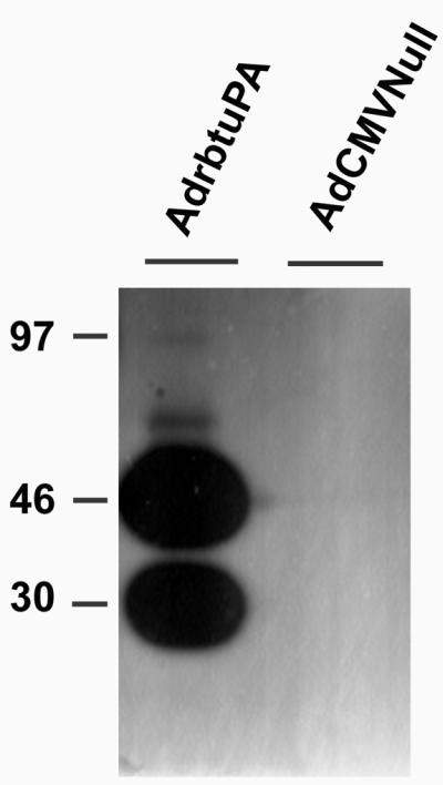
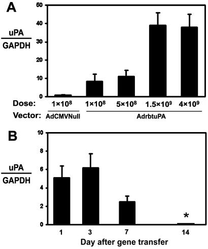
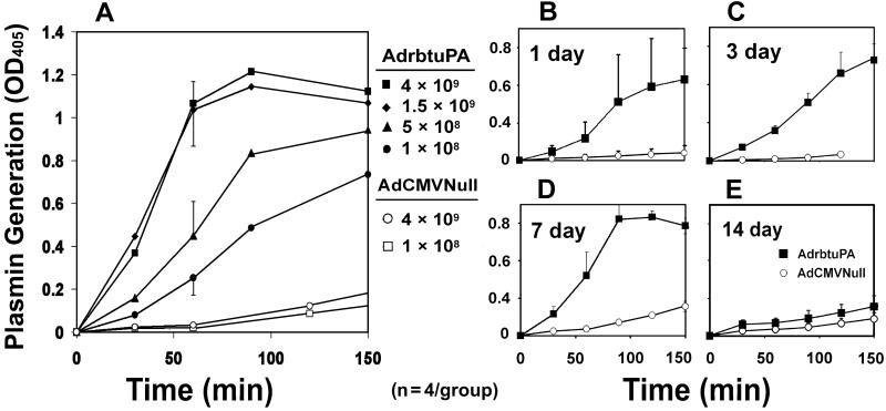
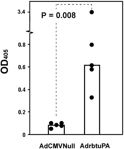
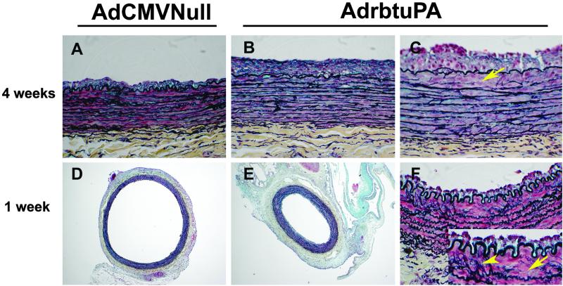
Overexpression of urokinase plasminogen activator (uPA) in endothelial cells can decrease intravascular thrombosis. However, expression of uPA is increased in atherosclerotic human arteries, which suggests that uPA might accelerate atherogenesis. To investigate whether elevated uPA expression accelerates atherogenesis, we cloned a rabbit uPA cDNA and expressed it in carotid arteries of cholesterol-fed rabbits. uPA gene transfer increased artery-wall uPA activity for at least 1 week, with a return to baseline by 2 weeks. One week after gene transfer, uPA-transduced arteries were constricted, with significantly smaller lumens and thicker walls, but no difference in intimal or medial mass. Two weeks after gene transfer, uPA- and control-transduced arteries were morphologically indistinguishable. By 4 weeks, however, uPA-transduced arteries had 70% larger intimas than control-transduced arteries (P < 0.01) and smaller lumens (P < 0.05). Intimal lesions appeared to be of similar composition in uPA- and control-transduced arteries, except that degradation of elastic laminae was evident in uPA-transduced arteries. These data suggest that elevated uPA expression in atherosclerotic arteries contributes to intimal growth and constrictive remodeling leading to lumen loss. Antagonists of uPA activity might, therefore, be useful in limiting intimal growth and preventing constrictive remodeling. Overexpression of uPA in endothelial cells to prevent intravascular thrombosis must be reconsidered, because this intervention could worsen underlying vascular disease.
Figures
Similar articles
-
Constriction of carotid arteries by urokinase-type plasminogen activator requires catalytic activity and is independent of NH(2)-terminal domains.Thromb Haemost. 2009 Nov;102(5):983-92. doi: 10.1160/TH09-03-0161. Thromb Haemost. 2009. PMID: 19888538 Free PMC article.
-
Improved vascular gene transfer with a helper-dependent adenoviral vector.Circulation. 2004 Sep 14;110(11):1484-91. doi: 10.1161/01.CIR.0000141574.78032.A9. Epub 2004 Aug 23. Circulation. 2004. PMID: 15326058
-
Macrophage-targeted overexpression of urokinase causes accelerated atherosclerosis, coronary artery occlusions, and premature death.Circulation. 2004 May 4;109(17):2129-35. doi: 10.1161/01.CIR.0000127369.24127.03. Epub 2004 Apr 19. Circulation. 2004. PMID: 15096455
-
Expression of Fas ligand in arteries of hypercholesterolemic rabbits accelerates atherosclerotic lesion formation.Arterioscler Thromb Vasc Biol. 2000 Feb;20(2):298-308. doi: 10.1161/01.atv.20.2.298. Arterioscler Thromb Vasc Biol. 2000. PMID: 10669624
-
Regulation of arterial remodeling and angiogenesis by urokinase-type plasminogen activator.Can J Physiol Pharmacol. 2009 Apr;87(4):231-51. doi: 10.1139/Y08-113. Can J Physiol Pharmacol. 2009. PMID: 19370078 Review.
Cited by
-
Expansive arterial remodeling: location, location, location.Arterioscler Thromb Vasc Biol. 2004 Apr;24(4):650-7. doi: 10.1161/01.ATV.0000120376.09047.fe. Epub 2004 Feb 5. Arterioscler Thromb Vasc Biol. 2004. PMID: 14764423 Free PMC article. Review.
-
In Vivo Gene Transfer to the Rabbit Common Carotid Artery Endothelium.J Vis Exp. 2018 May 6;(135):56982. doi: 10.3791/56982. J Vis Exp. 2018. PMID: 29782016 Free PMC article.
-
Improved animal models for testing gene therapy for atherosclerosis.Hum Gene Ther Methods. 2014 Apr;25(2):106-14. doi: 10.1089/hgtb.2013.199. Epub 2014 Feb 14. Hum Gene Ther Methods. 2014. PMID: 24528162 Free PMC article.
-
Helper-dependent adenovirus is superior to first-generation adenovirus for expressing transgenes in atherosclerosis-prone arteries.Arterioscler Thromb Vasc Biol. 2011 Jun;31(6):1317-25. doi: 10.1161/ATVBAHA.111.225516. Epub 2011 Mar 31. Arterioscler Thromb Vasc Biol. 2011. PMID: 21454808 Free PMC article.
-
Localization of cysteine protease, cathepsin S, to the surface of vascular smooth muscle cells by association with integrin alphanubeta3.Am J Pathol. 2006 Feb;168(2):685-94. doi: 10.2353/ajpath.2006.050295. Am J Pathol. 2006. PMID: 16436681 Free PMC article.
References
-
- Bachmann F. (2001) in Hemostasis and Thrombosis: Basic Principles and Clinical Practice, eds. Colman, R. W., Hirsh, J., Marder, V. J., Clowes, A. W. & George, J. N. (Lippincott, Philadelphia), pp. 275–320.
-
- Dichek D. A., Lee, S. W. & Nguyen, N. H. (1994) Blood 84, 504-516. - PubMed
-
- Dichek D. A., Anderson, J., Kelly, A. B., Hanson, S. R. & Harker, L. A. (1996) Circulation 93, 301-309. - PubMed
-
- Vassalli G. & Dichek, D. A. (1997) Cardiovasc. Res. 35, 459-469. - PubMed
-
- Kienast J., Padro, T., Steins, M., Li, C. X., Schmid, K. W., Hammel, D., Scheld, H. H. & van de Loo, J. C. (1998) Thromb. Haemostasis 79, 579-586. - PubMed
Publication types
MeSH terms
Substances
Associated data
- Actions
Grants and funding
LinkOut - more resources
Full Text Sources
Other Literature Sources
Molecular Biology Databases
Miscellaneous
