. 2002 Aug 6;99(16):10464-9.
doi: 10.1073/pnas.162301699. Epub 2002 Jul 29.

Neuregulin-1 promotes formation of the murine cardiac conduction system

Affiliations

Neuregulin-1 promotes formation of the murine cardiac conduction system

Stacey Rentschler et al. Proc Natl Acad Sci U S A. .

Abstract

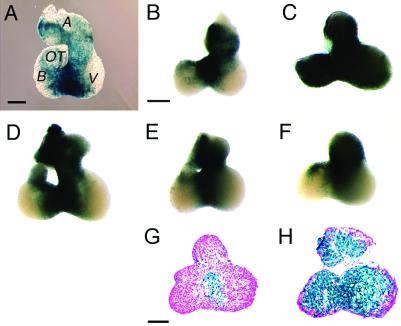
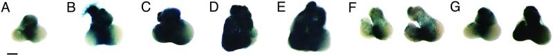
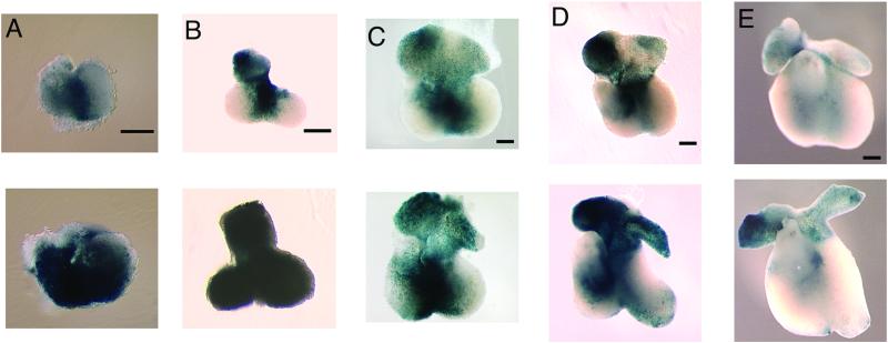
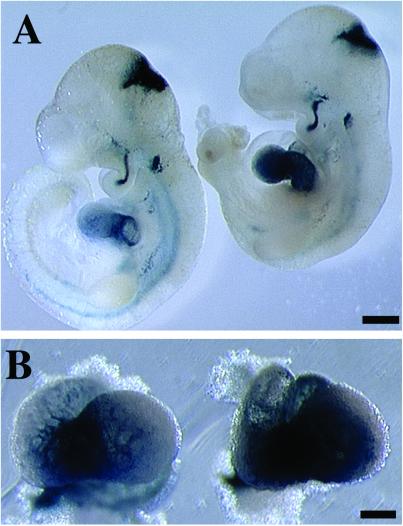
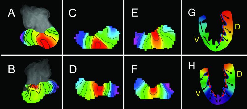
The cardiac conduction system is a network of cells responsible for the rhythmic and coordinated excitation of the heart. Components of the murine conduction system, including the peripheral Purkinje fibers, are morphologically indistinguishable from surrounding cardiomyocytes, and a paucity of molecular markers exists to identify these cells. The murine conduction system develops in close association with the endocardium. Using the recently identified CCS-lacZ line of reporter mice, in which lacZ expression delineates the embryonic and fully mature conduction system, we tested the ability of several endocardial-derived paracrine factors to convert contractile cardiomyocytes into conduction-system cells as measured by ectopic reporter gene expression in the heart. In this report we show that neuregulin-1, a growth and differentiation factor essential for ventricular trabeculation, is sufficient to induce ectopic expression of the lacZ conduction marker. This inductive effect of neuregulin-1 was restricted to a window of sensitivity between 8.5 and 10.5 days postcoitum. Using the whole mouse embryo culture system, neuregulin-1 was shown to regulate lacZ expression within the embryonic heart, whereas its expression in other tissues remained unaffected. We describe the electrical activation pattern of the 9.5-days postcoitum embryonic mouse heart and show that treatment with neuregulin-1 results in electrophysiological changes in the activation pattern consistent with a recruitment of cells to the conduction system. This study supports the hypothesis that endocardial-derived neuregulins may be the major endogenous ligands responsible for inducing murine embryonic cardiomyocytes to differentiate into cells of the conduction system.


Figures

Fig 1.
Fig 1.
NRG-1 converts 9.5-dpc embryonic cardiomyocytes to a CCS phenotype. X-Gal staining of a CCS-lacZ transgenic heart immediately after removal from a 9.5-dpc embryo (A). Highest lacZ expression can be seen in the area surrounding the bulboventricular junction in the presumptive “interventricular ring”, as well as in an area located on the right side of the common atrial chamber which may correspond to the developing sino-atrial node. CCS-lacZ hearts were cultured for 48 h in media alone (B), or in the presence of paracrine factors including a biologically active NRG-1 peptide (C), ET-1 (D), angiotensin II (E), or insulin-like growth factor-I (F), followed by X-Gal staining. G and H show a representative eosin-stained section from a control and NRG-1-treated heart, respectively. A, common atrium; V, ventricle; B, bulbus cordis; OT, outflow tract. [Bar = 0.2 mm (A); = 0.2 mm (BF); = 0.1 mm (G and H).]
Fig 2.
Fig 2.
Dose–response and time course of ectopic lacZ expression. CCS-lacZ hearts from 9.5-dpc embryos were cultured for 48 h with increasing concentrations of the NRG-1 EGF-like domain: (A) none, (B) 2.5 × 10⁻¹¹ M, (C) 2.5 × 10⁻¹⁰ M, (D) 2.5 × 10⁻⁹ M, or (E) 2.5 × 10⁻⁸ M, followed by X-Gal staining. CCS-lacZ hearts cultured for 24 h (F) or 48 h (G) in the absence (left heart) or presence of 2.5 × 10⁻⁹ M NRG-1 (right heart). [Bar = 0.2 mm (AG).]
Fig 3.
Fig 3.
Response to NRG-1 is developmentally regulated. CCS-lacZ hearts from (A) 8.5-dpc, (B) 9.5-dpc, (C) 10.5-dpc, (D) 11.5-dpc, and (E) 12.5-dpc embryos were cultured for 48 h in the absence (Upper) or presence of NRG-1 EGF-like domain (Lower) followed by X-Gal staining. Hearts were oriented with the atria located at the top of the picture for each stage except 8.5 dpc, where the atria are not visible. (Bars = 0.2 mm.)
Fig 4.
Fig 4.
NRG-1 induces ectopic CCS formation in treatment of whole embryos. CCS-lacZ embryos were harvested at 8–8.5 dpc, cultured in the absence (Left) or presence (Right) of NRG-1, and allowed to develop to 9.5–10.0 dpc. Extracardiac tissues that expressed lacZ were unaffected by NRG-1 treatment (A), whereas a higher magnification view of the ventricular areas of the hearts clearly shows ectopic lacZ expression within the heart (B). [Bar = 0.5 mm (A); = 0.2 mm (B).]
Fig 5.
Fig 5.
NRG-1 induces electrophysiological changes within embryonic hearts. Activation maps of 9.5-dpc wild-type hearts from either the ventral (A, C, E) or dorsal (B, D, F) surfaces are shown. Acutely dissociated hearts (A, B); hearts cultured for 24 h in the absence (C, D) or presence (E, F) of NRG-1. Activation progresses from red to purple, with isochrone lines drawn every 1 ms. Model depicting CCS-dependent activation of the ventricles in control (G) and NRG-1 (H) treated hearts. A sagittal view is shown with ventral (V) and dorsal (D) surfaces indicated. The CCS is indicated by the blue region along the subendocardial surface; activation sequence is indicated by colors progressing from red to purple. Red arrows indicate activation of the CCS; black arrows indicate activation of the working myocardium.

References

    1. Gourdie R. G., Mima, T., Thompson, R. P. & Mikawa, T. (1995) Development (Cambridge, U.K.) 121, 1423-1431. - PubMed
    1. Cheng G., Litchenberg, W. H., Cole, G. J., Mikawa, T., Thompson, R. P. & Gourdie, R. G. (1999) Development (Cambridge, U.K.) 126, 5041-5049. - PubMed
    1. Mikawa T. & Fischman, D. A. (1996) Annu. Rev. Physiol. 58, 509-521. - PubMed
    1. Yoshizumi M., Kurihara, H., Sugiyama, T., Takaku, F., Yanagisawa, M., Masaki, T. & Yazaki, Y. (1989) Biochem. Biophys. Res. Commun. 161, 859-864. - PubMed
    1. Gourdie R. G., Wei, Y., Kim, D., Klatt, S. C. & Mikawa, T. (1998) Proc. Natl. Acad. Sci. USA 95, 6815-6818. - PMC - PubMed

Publication types

LinkOut - more resources