. 2002 Aug 6;99(16):10330-4.
doi: 10.1073/pnas.162362899. Epub 2002 Jul 29.

Absolute requirement of spermidine for growth and cell cycle progression of fission yeast (Schizosaccharomyces pombe)

Affiliations

Absolute requirement of spermidine for growth and cell cycle progression of fission yeast (Schizosaccharomyces pombe)

Manas K Chattopadhyay et al. Proc Natl Acad Sci U S A. .

Abstract

Schizosaccharomyces pombe cells that cannot synthesize spermidine or spermine because of a deletion-insertion in the gene coding for S-adenosylmethionine decarboxylase (Deltaspe2) have an absolute requirement for spermidine for growth. Flow cytometry studies show that in the absence of spermidine an overall delay of the cell cycle progression occurs with some accumulation of cells in the G(1) phase; as little as 10(-6) M spermidine is sufficient to maintain normal cell cycle distribution and normal growth. Morphologically some of the spermidine-deprived cells become spherical at an early stage with little evidence of cell division. On further incubation in the spermidine-deprived medium, growth occurs in most of the cells, not by cell division but rather by cell elongation, with an abnormal distribution of the actin cytoskeleton, DNA (4', 6-diamidino-2-phenylindole staining), and calcofluor-staining moieties. More prolonged incubation in the spermidine-deficient medium leads to profound morphological changes including nuclear degeneration.


Figures

Fig 1.
Fig 1.
Growth curve of Δspe2 cells grown in the presence or absence of 10−6 M spermidine (Spd). The cultures were grown in 10−6 M spermidine and diluted into media containing no spermidine or 10−6 M spermidine, as described in Materials and Methods. The cultures were diluted into media with and without spermidine when necessary to keep the OD600 0.6–1.0. The optical densities presented have been corrected for these dilutions.
Fig 2.
Fig 2.
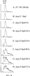
Histogram showing the FACS profile of spermidine (Spd)-supplemented and spermidine-depleted cells. Samples were obtained after growing the cells for different times in the polyamine-deficient medium (Fig. 1). Cells (5 × 10⁶) were harvested, fixed, and stained with Sybr Green I as detailed in Materials and Methods. The samples were then analyzed for DNA content by a Coulter Epics FACS (XL-MCL, Hialeah, FL).
Fig 3.
Fig 3.
The morphological features of Δspe2 cells during polyamine deprivation. The Δspe2 cells were grown as shown in Fig. 1; samples were harvested for various staining procedures and for differential interference-contrast optics (Nomarski) as described in Materials and Methods. (AD) Nomarski; (EH) nuclei staining by DAPI; (IL) actin and nuclei staining by rhodamine-phalloidin and DAPI, respectively; (MP) cell wall and septum staining by Calcofluor White M2R. Cells were observed under the fluorescence microscope and photographs were taken in a Zeiss Axiophot microscope equipped with a digital camera (Cool Snap HQ, Photometrics). (Bar = 10 μm.) (Insets) Magnified views showing the details of actin dots of the stained cells. Spd, spermidine; DIC, differential interference contrast microscopy.
Fig 4.
Fig 4.
Effect of different concentrations of added spermidine (Spd) on growth rate, FACS profile and DAPI staining. The Δspe2 cells were grown for 48 h in 10−6 M spermidine and diluted in different concentrations of spermidine in EMM medium. The growth rate, FACS analysis, and nuclear staining were performed when they were growing for 2–3 days in defined concentrations of spermidine. FACS analysis and DAPI staining were performed by using 70% ethanol-fixed cells. (Bar = 10 μm.)

References

    1. Tabor C. W. & Tabor, H. (1984) Annu. Rev. Biochem. 53, 749-790. - PubMed
    1. Pegg A. E. (1986) Biochem. J. 234, 249-262. - PMC - PubMed
    1. Cohen S. S., (1998) A Guide to the Polyamines (Oxford Univ. Press, New York), pp. 1–595.
    1. Balasundaram D., Tabor, C. W. & Tabor, H. (1991) Proc. Natl. Acad. Sci. USA 88, 5872-5876. - PMC - PubMed
    1. Balasundaram D., Dinman, J. D., Wickner, R. B., Tabor, C. W. & Tabor, H. (1994) Proc. Natl. Acad. Sci. USA 91, 172-176. - PMC - PubMed

MeSH terms

LinkOut - more resources