. 2002 Aug 1;22(15):6426-36.
doi: 10.1523/JNEUROSCI.22-15-06426.2002.

Recruitment of the kainate receptor subunit glutamate receptor 6 by cadherin/catenin complexes

Affiliations

Recruitment of the kainate receptor subunit glutamate receptor 6 by cadherin/catenin complexes

Françoise Coussen et al. J Neurosci. .

Abstract

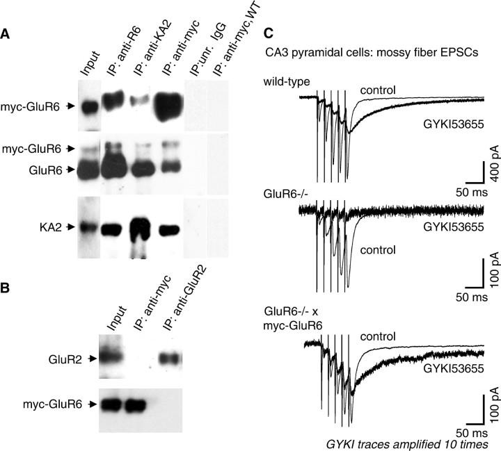
Kainate receptors modulate synaptic transmission by acting either at presynaptic or at postsynaptic sites. The precise localization of kainate receptors as well as the mechanisms of targeting and stabilization of these receptors in neurons are largely unknown. We have generated transgenic mice expressing the kainate receptor subunit glutamate receptor 6 (GluR6) bearing an extracellular myc epitope (myc-GluR6), in forebrain neurons, in which it assembles with endogenous kainate receptor subunits. In transgenic mice crossed with GluR6-deficient mice, myc-GluR6 efficiently rescues the missing subunit. Immunoprecipitation of transgenic brain extracts with anti-myc antibodies demonstrates an interaction with cadherins, beta-catenin, and p120 catenin, as well as with the associated proteins calcium calmodulin-dependent serine kinase and Velis, but not with alpha-catenin. In glutathione S-transferase-pulldown experiments, beta-catenin interacts, although indirectly, with the last 14 aa of GluR6. Transfected myc-GluR6 colocalizes with beta-catenin at cell-cell junctions in non-neuronal cells. Finally, activation of N-cadherins by ligand-covered latex beads recruits GluR6 to cadherin/catenin complexes. These results suggest an important role for cadherin/catenin complexes in the stabilization of kainate receptors at the synaptic membrane during synapse formation and remodeling.


Figures

Fig. 1.
Fig. 1.
Characterization of myc-GluR6 cDNA and transgenic mice. A, Functional expression of myc-GluR6 in HEK-293 cells. Cells were transiently transfected with myc-GluR6 cDNA. Whole-cell currents recorded in response to application of kainate (20 μm) in the presence of Con A are shown. Holding current, −70 mV. B, Surface expression of myc-GluR6 transiently transfected in COS-7 cells and cultured hippocampal neurons revealed with an anti-myc antibody. C, Schematic drawing of myc-GluR6 transgene construct. D, Expression of myc-GluR6 in selected brain regions for three transgenic mice families. Brain membrane extracts (15 μg) were analyzed in immunoblots probed with anti-myc (top) or anti-GluR6/7 (bottom) antibodies. Myc-GluR6 is differentially expressed in forebrain regions (hippocampus, striatum, and neocortex), but no expression is detected in the cerebellum. In wild-type mice (WT; right lane), no band is detected with the anti-myc antibody. Arrows indicate molecular weight markers.
Fig. 2.
Fig. 2.
Myc-GluR6 is associated with endogenous KAR subunits. A, Coimmunoprecipitation of myc-GluR6 with endogenous GluR6 and KA2 subunits. Triton X-100 brain extracts from transgenic mice were immunoprecipitated (IP) with anti-myc, anti-GluR6, or anti-KA2 antibodies. Immunoblot analysis was performed with the three antibodies. Control experiments (right lanes): immunoprecipitation of transgenic mouse brain extracts with an unrelated IgG (anti-GST antibody); immunoprecipitation of wild-type (WT) mouse brain extracts with an anti-myc antibody. B, Myc-GluR6 is not associated with GluR2 AMPA receptor subunit. Triton X-100 brain extracts from transgenic mice were immunoprecipitated with anti-myc or anti-GluR2 antibodies, and both proteins were analyzed by Western blot.C, Rescue of KAR-mediated synaptic currents in GluR6−/− mice by the myc-GluR6 transgene. Whole-cell patch-clamp recordings of CA3 pyramidal cells in hippocampal slices in the presence of antagonists of GABAA and NMDA receptors (10 μm bicuculline and 25 μmD-AP-5) are shown. Synaptic currents were evoked by a train of stimulations to mossy fibers. Blockade of AMPA receptors by the noncompetitive antagonist GYKI53655 revealed a slow synaptic component of small amplitude mediated by GluR6-containing KARs. The trace in the presence of GYKI was amplified 10-fold. The slow synaptic currents are not detected in GluR6−/− mice and are rescued in GluR6−/− mice mated with myc-GluR6 transgenic mice. Holding current, −70 mV. Calibration: 50 msec, 100 pA (except for wild type, 400 pA).
Fig. 3.
Fig. 3.
β-catenin associates with myc-GluR6.A, Coimmunoprecipitation of β-catenin and myc-GluR6. Brain extracts from transgenic or wild-type (WT;right lane) mouse brains (input, 1% of total brain extract) and anti-myc (20%), and anti-β-catenin (5%) immunoprecipitates (IP) were loaded on SDS-PAGE and probed with an anti-β-catenin antibody. B, Immunoprecipitation of β-catenin with antiglutamate receptor antibodies. The immunoprecipitate was immunoblotted with an anti-β-catenin antibody. The amount of β-catenin coimmunoprecipitated with anti-GluR6 or anti-KA2 antibodies represents, respectively, 2.5 and 16% of β-catenin immunoprecipitated with the anti-myc antibody. C, The amount of β-catenin associated with myc-GluR6 depends on the brain structure analyzed. Triton X-100 brain extracts (1.5 ml) from selected forebrain regions were immunoprecipitated with anti-myc antibodies and immunoblotted with anti-β-catenin antibody (10% of the immunoprecipitate fractions). Numbers correspond to the fractions of β-catenin immunoprecipitated for a same amount of total GluR6 loaded on the gel (GluR6 plus myc-GluR6) relative to the maximum amount of β-catenin immunoprecipitated in the corresponding experiment (IP myc lane for B and Hippocampus lanefor C). D, Immunoprecipitation of myc-GluR6 with anti β-catenin antibodies. E, Immunoprecipitation of p120 catenin and cadherin with anti-myc antibodies but not with anti-GluR6 or anti-KA2 antibodies.F, α-Catenin was not immunoprecipitated with an anti-myc antibody. For each immunoprecipitation (D–F), Triton X-100 brain extracts (1.5 ml) were incubated with the corresponding antibodies and submitted to immunoprecipitation. Input: 1% of the total brain extract was loaded; when the same antibody was used for immunoprecipitation and immunoblotting, 5% of the sample was loaded; when a different antibody was used for immunoprecipitation and immunoblotting, 20% of the sample was loaded. IB, Antibody used for immunoblotting.
Fig. 4.
Fig. 4.
Myc-GluR6 colocalizes with endogenous β-catenin and synaptophysin in transfected hippocampal neurons. Myc-GluR6 was transfected in cultured hippocampal neurons at 12 d in vitro. One day later, neurons were labeled in vivo (20 min at 20°C) with an anti-myc antibody, fixed, permeabilized, and then labeled for β-catenin (top panels) or synaptophysin (bottom panels). Labeling for anti-myc antibody is shown in red, and labeling for anti-β-catenin or anti-synaptophysin antibodies is shown in green. Arrows indicate colocalization between the two proteins. Scale bars are indicated. The boxed areas from the top panels are enlarged at the bottom of the figures.
Fig. 5.
Fig. 5.
CASK and Velis associate with myc-GluR6.A, Coimmunoprecipitation of CASK with β-catenin and Velis. Triton X-100 brain extracts (1.5 ml) were incubated with the corresponding antibodies, and 20% of the immunoprecipitate (IP) samples were immunoblotted with an anti-CASK antibody. Rightlane, Control experiment using an unrelated IgG (anti-GST antibody). B, Coimmunoprecipitation of Velis and myc-GluR6. C, Coimmunoprecipitation of CASK and myc-GluR6. D, Coimmunoprecipitation of myc-GluR6 by anti-Veli and anti-CASK antibodies. B–D, In the input lane, 1% of the total brain extract was loaded; when the same antibody was used for immunoprecipitation and immunoblotting, 5% of the sample was loaded; when a different antibody was used for immunoprecipitation and immunoblotting, 25% of the sample was loaded. B, C,Right lane corresponds to a control immunoprecipitation on wild-type (WT) brain extracts using an anti-myc antibody. IB, Antibody used for immunoblotting.
Fig. 6.
Fig. 6.
C-terminal domain of GluR6 is necessary for β-catenin and CASK binding. A–C, GST-pulldown of PSD-95, CASK, and β-catenin. Brain supernatant was submitted for binding to GST and GST proteins fused to total GluR6 C terminal (R6Ctotal) and to GluR6 deleted of the last 4 (R6C Δ4) or 14 (R6C Δ14) aa. Twenty percent of the samples were immunoblotted and probed for PSD-95 (A), CASK (B), β-catenin (C), and myc-GluR6 (D). E, Indirect binding of β-catenin to the C-terminal domain of GluR6. β-catenin expressed in bacteria was submitted for binding to the GST-cytoplasmic domain of N-cadherin (N-Cad), GST-GluR6 total C terminal, and GST-GluR6Δ14. As a control, PSD-95 (PDZ domain 1–2) expressed in bacteria was submitted for binding to GST-GluR6 total C terminal. SDS gels were submitted for blotting with GST, β-catenin, or PSD-family antibodies as indicated. IB, Antibody used for immunoblotting.
Fig. 7.
Fig. 7.
Endogenous β-catenin coimmunoprecipitates and colocalizes with myc-GluR6 in transfected COS-7 cells. Myc-GluR6 or myc-GluR6Δ14 were transfected with or without PSD-95 in COS-7 cells. After 1 d, cells were extracted, and Triton X-100 supernatants were immunoprecipitated with an anti-myc antibody. A, Immunoprecipitates (IP) were immunoblotted with anti-myc, anti-PSD-95, or anti-β-catenin antibodies. Endogenous β-catenin coimmunoprecipitates with myc-GluR6 in transfected COS-7 cells. B, Immunocytochemistry of COS-7 cells transfected with myc-GluR6, myc-GluR6Δ14, or myc-GluR6 plus PSD-95 with an anti-myc antibody (surface labeling) and an anti-β-catenin antibody (intracellular labeling). Myc-GluR6, but not myc-GluR6Δ14, colocalizes with β-catenin at cell–cell junctions. Expression of PSD-95 decreases localization of myc-GluR6 at the junction of the cells.
Fig. 8.
Fig. 8.
Myc-GluR6 redistribution follows the distribution of β-catenin. C2 cells were transfected with myc-GluR6 (A, C, D) or myc-GluR6Δ14 (B). Twenty-four hours later, Ncad-Fc- (A, B) or anti-myc- (C, D) coated beads were incubated with cells for 45 min. Cells were fixed, permeabilized, and probed with anti-myc tag or anti-β-catenin antibodies. Activation of cadherin induces recruitment of myc-GluR6 under the bead (A). There is no recruitment of myc-GluR6Δ14 by Ncad-Fc-coated beads (B). Anti-myc antibody-coated beads induce a strong recruitment of myc-GluR6 at the bead–cell contact (C). However, recruitment of myc-GluR6 is not followed by recruitment of β-catenin under the beads (D).

References

    1. Benson DL, Tanaka H. N-cadherin redistribution during synaptogenesis in hippocampal neurons. J Neurosci. 1998;18:6892–6904. - PMC - PubMed
    1. Bettler B, Mulle C. AMPA and kainate receptors. Neuropharmacology. 1995;34:123–139. - PubMed
    1. Bischoff S, Barhanin J, Bettler B, Mulle C, Heinemann S. Spatial distribution of kainate receptor subunit mRNA in the mouse basal ganglia and ventral mesencephalon. J Comp Neurol. 1997;379:541–562. - PubMed
    1. Bortolotto ZA, Clarke VR, Delany CM, Parry MC, Smolders I, Vignes M, Ho KH, Miu P, Brinton BT, Fantaske R, Ogden A, Gates M, Ornstein PL, Lodge D, Bleakman D, Collingridge GL. Kainate receptors are involved in synaptic plasticity. Nature. 1999;402:297–301. - PubMed
    1. Bozdagi O, Shan W, Tanaka H, Benson DL, Huntley GW. Increasing numbers of synaptic puncta during late-phase LTP: N-cadherin is synthesized, recruited to synaptic sites, and required for potentiation. Neuron. 2000;28:245–259. - PubMed

Publication types

MeSH terms

Substances