. 2002 Aug 1;22(15):6437-46.
doi: 10.1523/JNEUROSCI.22-15-06437.2002.

Distribution of a lysosomal enzyme in the adult brain by axonal transport and by cells of the rostral migratory stream

Affiliations

Distribution of a lysosomal enzyme in the adult brain by axonal transport and by cells of the rostral migratory stream

Marco A Passini et al. J Neurosci. .

Abstract

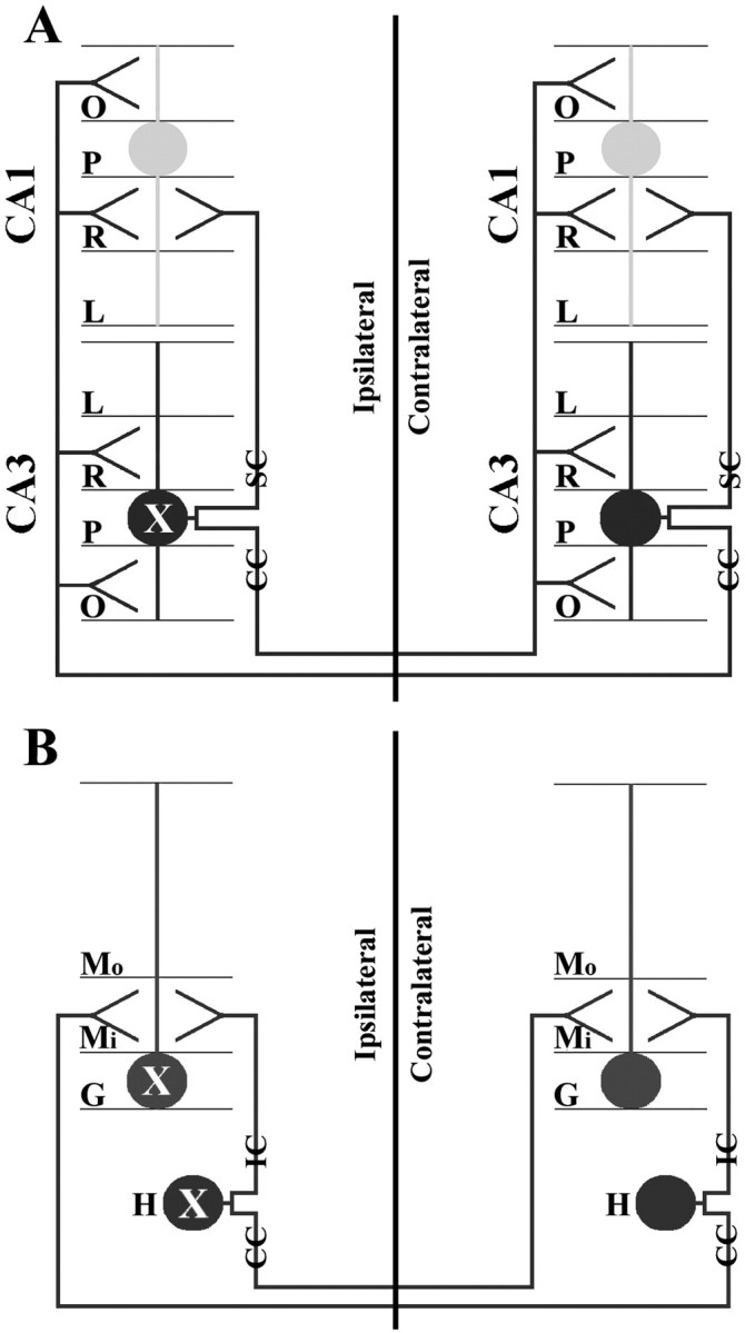
A portion of the lysosomal enzymes produced by cells is secreted, diffuses through extracellular spaces, and can be taken up by distal cells via mannose-6-phosphate receptor-mediated endocytosis. This provides the basis for treating lysosomal storage diseases, many of which affect the CNS. Normal enzyme secreted from a cluster of genetically corrected cells has been shown to reverse storage lesions in a zone of surrounding brain tissue in mouse disease models. However, low levels of enzyme activity and reduction of storage lesions also have been observed at sites in the brain that may not be explained by a contiguous gradient of secreted enzyme diffusing away from the genetically corrected cells. No direct evidence for alternative mechanisms of enzyme transport has been shown, and little is understood about the intracellular movement of lysosomal enzymes in neurons. We investigated whether axonal transport could occur, by expressing an eukaryotic lysosomal enzyme that can be visualized in tissue sections (beta-glucuronidase) in brain structures that have defined axonal connections to other structures. This resulted in the transfer of enzyme to, and a reversal of storage lesions in, neurons that project to the gene expression site, but not in nearby structures that would have been corrected if the effect had been mediated by diffusion. In addition, transduction of cells in the subventricular zone resulted in the uptake of beta-glucuronidase by cells entering the rostral migratory stream. Gene transfer to specific neuronal circuits or cells in migratory pathways may facilitate delivery to the global brain lesions found in these disorders.


Figures

Fig. 1.
Fig. 1.
Comparison of AAV2-HβH-injected (A–C) and AAV2-HβH-uninjected (D–F) brains in the C3H/HeOuJmouse strain. Enzyme histochemistry (A, D) and in situ hybridization with the antisense riboprobe (ISH-antisense;B, E) detected many positive cells at 1 month PI, but did not detect positive cells in age-matched uninjected brains.In situ hybridization with the sense riboprobe (negative control) produced little positive signal in the injected brain (C) and no positive signal in the uninjected brain (F). CP, Caudate putamen;CTX, somatosensory cortex; EC, external capsule. Scale bar, 200 μm.
Fig. 2.
Fig. 2.
Comparison between AAV2-CVβ and AAV2-HβH in the hippocampus at 1 month PI (A–F) and 3 months PI (G–M). Shown are ISH-antisense (A–C, G–I) and enzyme histochemistry (D–F, J–M). The number of mRNA- and enzyme-positive cells decreased between 1 month PI (A, D) and 3 months PI (G, J) with AAV2-CVβ. In contrast, the number of cells expressing GUSB mRNA did not change between 1 month (B) and 3 months (H) with AAV2-HβH. In addition, a substantial increase in the number of enzyme-positive cells was detected at 3 months (K) compared with 1 month (E) with AAV2-HβH. Enzyme-positive cells were clearly present in regions of the ipsilateral hippocampus that were negative for gene expression (K). There was a conspicuous absence of enzyme staining in the CA1–CA3 lacunosum moleculare layers at 3 months (asterisk inK). The number of enzyme-positive cells in the contralateral hemisphere was also greater at 3 months (L) compared with 1 month (F) with AAV2-HβH. At 1 month the enzyme-positive cells were restricted to the inner molecular layer of the dentate gyrus (F, arrow). This was verified by the staining of an enzyme-positive section with 0.1% cresyl violet, which labeled the dentate granule cell layer, but not the dentate molecular layer (small box, F). At 3 months the pattern of enzyme-positive cells extended to include other regions in the contralateral hippocampus, particularly the oriens and radiatum layers of CA1–CA3 (L). A high-magnification image of a cresyl violet-stained section demonstrated that enzymatic activity was maintained in the inner molecular layer of the dentate gyrus (small box, L). Transduced cells were not detected in the uninjected hemisphere at either time point with AAV2-HβH-injected brains (C, I). An entire brain section at 3 months PI demonstrated how well enzymatic activity was confined to the hippocampus, although a small number of positive cells could be seen in the overlying cortex (M). CA1p, CA1 pyramidal cell layer; CA3p, CA3 pyramidal cell layer;G, upper and lower blades of the dentate granule cell layer; H, hilus; L, stratum lacunosum moleculare; Mi, inner molecular layer of the dentate gyrus; Mo, outer molecular layer of the dentate gyrus;O, stratum oriens; R, stratum radiatum. Scale bars: A–L, 500 μm; M, 1000 μm.
Fig. 3.
Fig. 3.
Long-term enzymatic activity and expression in the hippocampal commissure and hippocampus. Shown are enzyme histochemistry (A, B, G–I) and ISH-antisense (C–F) in the hippocampal commissure (A–C, I), ipsilateral hippocampus (D–G), and contralateral hippocampus (H). Unilateral injections of AAV2-HβH into the ventral hippocampus resulted in undetectable levels of enzyme staining at 1 month PI (A) but detectable levels at 3 months PI (B) in the hippocampal commissure. Although a small number of cell bodies were labeled, the majority of staining was present in a diffuse, linear pattern (B). Vector-encoded human GUSB mRNA was not present in a corresponding section at 3 months (C). Robust transduction was observed in the ventral hippocampus in all three mice at 18 months PI (D–F). In mouse 1 the GUSB-expressing cells were abundant in the CA3 pyramidal cell layer and in the ventral blade of the dentate GCL (D). In mouse 2 transduced cells were present in the pyramidal layer of CA2 and CA3 (E). In mouse 3 a large number of GUSB-expressing cells were detected in the CA3 pyramidal cell layer and in the GCL and hilus of the dentate gyrus (F). As represented by mouse 1, widespread enzyme-positive cells were detected in the injected (G) and uninjected (H) hemispheres of all three mice. The most robust enzyme staining in the uninjected hemisphere was detected in CA1–CA3 (H). Other regions, such as the inner molecular layer of the dentate gyrus, were also positive for enzymatic activity (H). A larger area of diffuse enzyme staining was observed in the hippocampal commissure at 18 months (I) compared with 1 month (A) and 3 months (B). Scale bars: A–D, G, H, 500 μm; E, F, I, 250 μm.
Fig. 4.
Fig. 4.
Reversal of pathology in the MPS VII brains after unilateral injection of AAV2-HβH into the ventral hippocampus at 3 months PI. Shown are toluidine blue-stained plastic sections in uninjected (A, D) and injected (B, C, E, F) mice. Lysosomal storage vacuoles were visible in the uninjected GCL and hilus of the dentate gyrus (A) but were cleared in the ipsilateral (B) and contralateral (C) hemispheres of injected brains. Lysosomal storage vacuoles, which were present in the entire CA3 pyramidal cell layer in uninjected mice (D), were reversed in both the ipsilateral (E) and contralateral (F) hemispheres of injected brains.Arrows point to lysosomal storage vacuoles. Scale bar, 50 μm.
Fig. 5.
Fig. 5.
Axonal transport in the nigrostriatal (A–D) and septohippocampal (E–H) systems after AAV2-HβH vector injection. Shown are enzyme histochemistry (A, C, E–G) and ISH-antisense (B, D, H). Injections into the striatum resulted in many enzyme- and mRNA-positive cells in this structure at 18 months PI (A, B). Enzyme-positive cells were present in the substantia nigra after the striatal injections (C). However, the entire substantia nigra was negative for gene expression (D). After injection of the ventral hippocampus, enzyme-positive cells were undetectable in the septum at 1 month (E) but were detectable at 3 months (F) and 18 months (G). Enzyme-positive cells in the septum were observed in the medial nucleus and in both dorsolateral nuclei, but not in either ventrolateral nucleus (F, G). ISH-antisense did not produce positive cells in the septum at 18 months (H). Scale bars: A–D, 250 μm; E–H, 500 μm.
Fig. 6.
Fig. 6.
Correction of storage in the septum after hippocampal injections. Shown are toluidine blue-stained plastic sections of the septum (A–D) and other brain structures (E–H). Storage vacuoles were present in many cells of the uninjected MPS VII medial (A) and lateral (C) septa. Reversal of pathology was observed in neurons and glial cells of the medial (B) and lateral (D) septum 3 months after AAV2-HβH vector injection of the MPS VII ventral hippocampus. These injections did not correct storage lesions in other brain structures, as illustrated by the neocortex (E), piriform cortex (F), caudate putamen (G), and thalamus (H). Arrows point to lysosomal storage vacuoles. Scale bar, 40 μm.
Fig. 7.
Fig. 7.
Transport of GUSB in the rostral migratory stream. Shown are enzyme histochemistry (A–C, G–I) and ISH-antisense (D–F, J–L) in coronal sections of the subventricular zone (A, D, G, J), the rostral migratory stream (B, E), and the main olfactory bulb (C, F, H, I, K, L) at 1 month PI (A–F) and 18 months PI (G–L). Scale bars: A, D, 500 μm; C, F, 400 μm; G, H, J, K, 250 μm; B, E, I, L, 60 μm.
Fig. 8.
Fig. 8.
Diagram showing the symmetrical circuitry of the intrahippocampal system. CA1 and CA3 pyramidal cells have dendrites that extend into the oriens, radiatum, and lacunosum layers (A). CA3 pyramidal cells send commissural collaterals to the contralateral CA1–CA3 radiatum and oriens layers and send Schaffer collaterals to the ipsilateral CA1 radiatum layer (A). Dentate granule cells have dendrites that extend into the inner and outer molecular layers (B). Hilar neurons of the dentate gyrus send ipsilateral and commissural collaterals to the inner one-third molecular layer and synapse with dentate granule cells (B). According to these connections the GUSB transport to the contralateral side probably occurred by secretion of GUSB into the ipsilateral synapse by the transduced cell (labeled asX), followed by uptake by the axonal termini (or terminus) of the contralateral cell and subsequent retrograde transport via the commissural axon to the soma. Although anterograde projections occur, it is not thought that lysosomes move to the synaptic terminus (Walkley, 1998). CC, Commissural collateral;IC, ipsilateral collateral; P, pyramidal cell layer; SC, Schaffer collateral; see Figure 2 for additional abbreviations.
Fig. 9.
Fig. 9.
GUSB movement throughout the circuit of the septohippocampal system. Only one hemisphere of the ventral hippocampus is shown. Neurons from the medial septum project onto CA3 pyramidal and dentate hilar cells, whereas CA3 pyramidal cells project onto the lateral septal nuclei of both hemispheres (Swanson, 1977). Retrograde transport in axons of the medial septum is the likely mechanism for GUSB movement in this circuit. Because neurons of the lateral septum also project onto the medial septum, the enzyme pattern and reversal of pathology in the lateral septum probably occur by retrograde transport via a second axon (second-order neuron). CA3p, CA3 pyramidal cell layer; H, hilus; LS, lateral septum; MS, medial septum.

Similar articles

Cited by

References

    1. Achord D, Brot F, Gonzalez-Noriega A, Sly W, Stahl P. Human β-glucuronidase. II. Fate of infused human placental β-glucuronidase in the rat. Pediatr Res. 1977;11:816–822. - PubMed
    1. Alexander IE, Russell DW, Spence AM, Miller AD. Effects of gamma irradiation on the transduction of dividing and nondividing cells in brain and muscle of rats by adeno-associated virus vectors. Hum Gene Ther. 1996;7:841–850. - PubMed
    1. Altman J. Autoradiographic and histological studies of postnatal neurogenesis. IV. Cell proliferation and migration in the anterior forebrain, with special reference to persisting neurogenesis in the olfactory bulb. J Comp Neurol. 1969;137:433–457. - PubMed
    1. Amaral DG, Witter MP. The three-dimensional organization of the hippocampal formation: a review of anatomical data. Neuroscience. 1989;31:571–591. - PubMed
    1. Barthel LK, Raymond PA. In situ hybridization studies of retinal neurons. Methods Enzymol. 2000;316:579–590. - PubMed

Publication types

MeSH terms

LinkOut - more resources