. 2002 Aug 1;22(15):6447-57.
doi: 10.1523/JNEUROSCI.22-15-06447.2002.

Active calcium accumulation underlies severe weakness in a panel of mice with slow-channel syndrome

Affiliations

Active calcium accumulation underlies severe weakness in a panel of mice with slow-channel syndrome

Christopher M Gomez et al. J Neurosci. .

Abstract

Mutations affecting the gating and channel properties of ionotropic neurotransmitter receptors in some hereditary epilepsies, in familial hyperekplexia, and the slow-channel congenital myasthenic syndrome (SCCMS) may perturb the kinetics of synaptic currents, leading to significant clinical consequences. Although at least 12 acetylcholine receptor (AChR) mutations have been identified in the SCCMS, the altered channel properties critical for disease pathogenesis in the SCCMS have not been identified. To approach this question, we investigated the effect of different AChR subunit mutations on muscle weakness and the function and viability of neuromuscular synapses in transgenic mice. Targeted expression of distinct mutant AChR subunits in skeletal muscle prolonged the decay phases of the miniature endplate currents (MEPCs) over a broad range. In addition, both muscle strength and the amplitude of MEPCs were lower in transgenic lines with greater MEPC duration. SCCMS is associated with calcium overload of the neuromuscular junctional sarcoplasm. We found that the extent of calcium overload of motor endplates in the panel of transgenic mice was influenced by the relative permeability of the mutant AChRs to calcium, on the duration of MEPCs, and on neuromuscular activity. Finally, severe degenerative changes at the motor endplate (endplate myopathy) were apparent by electron microscopy in transgenic lines that displayed the greatest activity-dependent calcium overload. These studies demonstrate the importance of control of the kinetics of AChR channel gating for the function and viability of the neuromuscular junction.


Figures
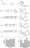
Fig. 1.
Fig. 1.
In vitro kinetic and permeation properties of a panel of mutant mouse AChRs used in transgenic mice.A, Single-channel recordings (left) and open duration histograms (right) for mouse wild-type and AChR mutants expressed in Xenopus oocytes. Recordings were performed at −100 mV and 2.0 μm ACh. Marquardt least squares fitting in pClamp 6 allowed resolution of two open components for each mutant (Table 1). B, Single-channel conductance obtained from oocyte patch-clamp recordings. There is no significant difference between the conductance for sodium among any of the mutants. C, Permeability of wild-type and mutant AChRs to calcium relative to sodium. The relative calcium permeability of αL251T (n = 17) AChRs is ∼33% greater than wild-type AChRs (n = 19;p < 0.005). The relative calcium permeability of δS262T AChRs (n = 20) is ∼67% less than wild type (p < 0.001).
Fig. 2.
Fig. 2.
Analysis of transgene mRNA expression. Expression of the mutant AChR mRNA in the panel of transgenic mice was comparable between several lines and well above that of endogenous mouse AChR subunit genes. A, Matched expression of AChR subunit transgenes. Top panel, Ten micrograms of total muscle RNA per mouse were hybridized with common sequence probe for all transgenes. Exposure was 4 d. Bottom panel, Same blot rehybridized with MCK cDNA to ensure uniform loading. Exposure was 4 hr. Lane 1, δS262T, line 40; lane 2, αC418W, line 42; lane 3, αL251T, line 39;lane 4, αL251T, line 36; lane 5, εL269F, line 14; lane 6, εL269F, line 5; lane 7, control muscle. B, Overexpression of mutant α subunits. Top panel, Total muscle RNA probed with mouse α subunit cDNA. Exposure was 5 d. Endogenous α subunit mRNA is undetectable at this explosure time. Lane 1, αC418W, line 42; lane 2, αL251T, line 39;lane 3, αL251T, line 36; lane 4, control muscle. Bottom panel, Same blot reprobed with MCK cDNA. Exposure was 4 hr. Size markers are in kilobases.
Fig. 3.
Fig. 3.
Miniature endplate current traces from individual diaphragm muscle fibers of wild type (A), δS262T (B), αC418W (C), αL251T (D), and εL269F (E). The prolonged and prominent bi-exponential decay phases correlate with the open times of the mutant channels expressed in the mice (Table 1). A, Average rise, 0.42 ± 0.47 msec; τs, 1.34 msec; amplitude (Amp), 4.15 ± 1.16 nA. B, Average rise, 0.32 ± 0.09 msec; τs = 2.34 msec; Amp, 2.69 ± 0.47 nA. C, Average rise, 0.25; τ1 = 1.52, τ2 = 9.89 msec; Amp, 3.48 nA. D, Average rise, 0.18 ± 0.14 msec; τ1 = 0.72 msec, τ2 = 8.37 msec; Amp, 3.31 ± 0.65 nA. E, Average rise, 0.37 ± 0.33 msec; τ1 = 0.17 msec, τ2 = 15.87 msec; Amp, 2.58 nA. Calibration: 1 nA, 15 msec.
Fig. 4.
Fig. 4.
Transgene-specific neuromuscular weakness correlates with MEPC current kinetics in slow-channel transgenic mice.A, Slow-channel mice exhibit a broad range of muscle weakness (left axis, dark bars). Strength testing was performed using a wire-hang paradigm (see Materials and Methods). Results are expressed as percentage of perfect score. The mean scores of αC418W (p < 0.025;n = 5), αL251T (p < 0.015; n = 4), and εL269F (p<<0.001;n = 8), but not that of δS262F (p = 0.5; n = 4), were significantly worse than age-matched control mice (n = 25). Performance by εL269F mice was also significantly poorer than αC418W mice (p< 0.02). Slow-channel mice have diminished miniature endplate current (MEPC) amplitudes (right axis,light bars). MEPC amplitudes (nanoamps) were recorded during voltage-clamp analysis of excised diaphragms of control (n = 5 mice) or δS262F (n = 4), αC418W (n = 7), αL251T (n = 6), or εL269F (n = 8) transgenic mice. The mean MEPC amplitude was significantly lower than control mice in εL269F transgenic mice (p< 0.025). B, εL269F (n = 7) transgenic mice have reduced endplate AChRs compared with control (p < 0.025; n = 4), as determined by 125I α−BT binding (see Materials and Methods). C, Plot of strength performance versus the slow decay time constant (τ2) of bi-exponential MEPCs for each transgenic mouse line. For this plot the value for τs was used for wild type because no τ2value was resolved for wild-type mice. D, Plot of strength performance versus the log of MEPC area as expressed in picocoulombs. Mouse lines for C and D:open circle, wild type; filled square, δS262; filled triangle, αC418W; filled diamond, αL251T; filled circle, εL269F.
Fig. 5.
Fig. 5.
Calcium accumulation at motor endplates differs with transgenic expression of distinct AChR mutations.A, Sections of forelimb flexor muscles from resting δS262T, αC418W, αL251T, and εL269F mice demonstrate differences in degree of GBHA staining for different transgenic lines. Sections in the left panels were stained for cholinesterase to localize endplates (cholinesterase is brown) (Namba et al., 1967) and counterstained with hematoxylin and eosin. The adjacent serial sections in the right panels were stained for glyoxal bis 2 hydroxyanil (Evans, 1974) (GBHA, dark red or black) and methylene blue. There is no GBHA staining at control (data not shown) or δS262T endplates. Scale bar, 25 μm. B, Calcium accumulation at motor endplates differs according to mutation and increases with exercise. The proportions of motor endplates stained histochemically with GBHA in αC418W, αL251T, and εL269F mice at rest or after exercise is displayed in vertical barslabeled below. Dark bars indicate the lines that develop endplate myopathy (Fig. 6). At rest, no GBHA stain was detected at control or δS262T endplates, whereas 0.47% were labeled in αC418W mice, 2.8% in αL251T, and 19% in εL269F mice. The proportion of Ca2+-overloaded endplates detected in resting αL251T mice (n = 6) was slightly greater than that in αC418W mice (0.47%; p = 0.08;n = 6). The proportion of GBHA-stained endplates in resting εL269F transgenic mice was significantly greater than that of the other resting mice (p ≪ 0.001;n = 5). After exercise (see Materials and Methods), there was still no GBHA staining at motor endplates or elsewhere in muscle fibers of wild-type or δS262T transgenic mice, whereas marked increases in GBHA staining were seen in the other three transgenic lines after exercise. In exercised αC418W transgenic mice (n = 5), the proportion of endplates stained for GBHA increased to 3.9%, an eightfold increase (p < 0.01). In αL251T transgenic mice (n = 4) and εL269F transgenic mice (n = 4), the proportion of GBHA-stained endplates increased to 26% (10-fold; p < 0.002) and 33% (1.7-fold; p < 0.04), respectively. The proportions of Ca2+-overloaded endplates after exercise in αL251T transgenic mice and εL269F transgenic mice were significantly greater than the Ca2+-overloaded endplates in exercised αC418W transgenic mice (p < 0.01).
Fig. 6.
Fig. 6.
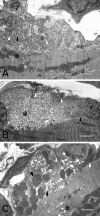
Endplate myopathy occurs in slow-channel transgenic mice with severe calcium overload. A, Neuromuscular junction from a 2-month-old αC418W mouse showing essentially normal ultrastructure of nerve terminus (white arrowheads), postsynaptic folds (black arrow), and junctional sarcoplasm (asterisk).B, Neuromuscular junction from 2-month-old αL251T mouse. The junctional sarcoplasm is filled with myriads of vacuole-like structures ranging in size from 0.05 to 1 μm (asterisk). Vacuoles are empty or filled with fluffy or granular material. Profiles of nuclei are present at either side of the accumulation of vacuoles. One nucleus appears severely degenerated (black arrow). In B and Cthe secondary synaptic folds and clefts are absent, and the nerve terminals (white arrowheads) are small and barely recognizable at the outer surface of the bulging postsynaptic regions.C, Neuromuscular junction from a εL269F mouse at 2 months. Vacuoles fill the junctional sarcoplasm and are present within the underlying sarcomeres (asterisk). The subsynaptic mitochondria are abnormally enlarged compared with those in the nerve terminus. Some show accumulations of dark, dense granules consistent with calcium (data not shown). Others contain multiple clear inclusions (black arrowhead). Some mitochondria are pathologically dilated (black arrow). One subsynaptic nucleus has normal ultrastructure. Scale bars: A, 0.9 μm;B, 4.1 μm; C, 1.5 μm.

Similar articles

Cited by

References

    1. Bertrand D, Galzi JL, Devillers TA, Bertrand S, Changeux JP. Mutations at two distinct sites within the channel domain M2 alter calcium permeability of neuronal alpha 7 nicotinic receptor. Proc Natl Acad Sci USA. 1993;90:6971–6975. - PMC - PubMed
    1. Bhattacharyya BJ, Day J, Gundeck JE, Leonard S, Wollmann RW, Gomez C. Desensitization of mutant acetylcholine receptors in transgenic mice reduces the amplitude of synaptic currents. Synapse. 1997;27:367–377. - PubMed
    1. Bodensteiner JB, Engel AG. Intracellular calcium accumulation in Duchenne dystrophy and other myopathies: a study of 567,000 muscle fibers in 114 biopsies. Neurology. 1978;28:439–446. - PubMed
    1. Boulter J, Luyten W, Evans K, Mason P, Ballivet M, Goldman D, Stengelin S, Martin G, Heinemann S, Patrick J. Isolation of a clone coding for the α-subunit of a mouse acetylcholine receptor. J Neurosci. 1985;5:2545–2552. - PMC - PubMed
    1. Brenner HR, Witzemann V, Sakmann B. Imprinting of acetylcholine receptor messenger RNA accumulation in mammalian neuromuscular synapses. Nature. 1990;344:544–547. - PubMed

Publication types

MeSH terms