. 2002 Aug 1;22(15):6578-86.
doi: 10.1523/JNEUROSCI.22-15-06578.2002.

Ceruloplasmin regulates iron levels in the CNS and prevents free radical injury

Affiliations

Ceruloplasmin regulates iron levels in the CNS and prevents free radical injury

Bharatkumar N Patel et al. J Neurosci. .

Abstract

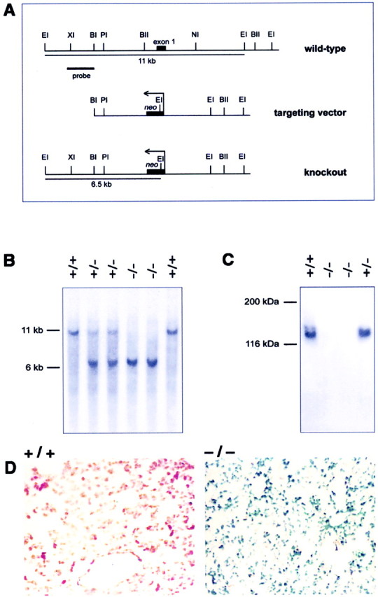
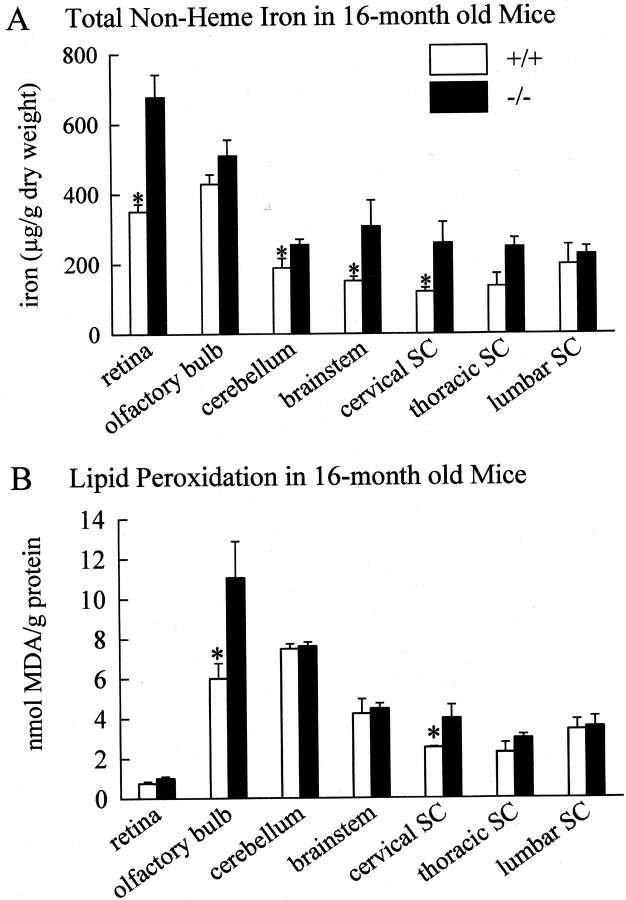
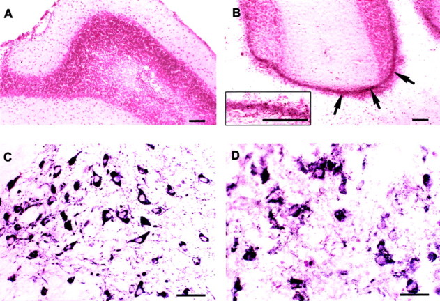
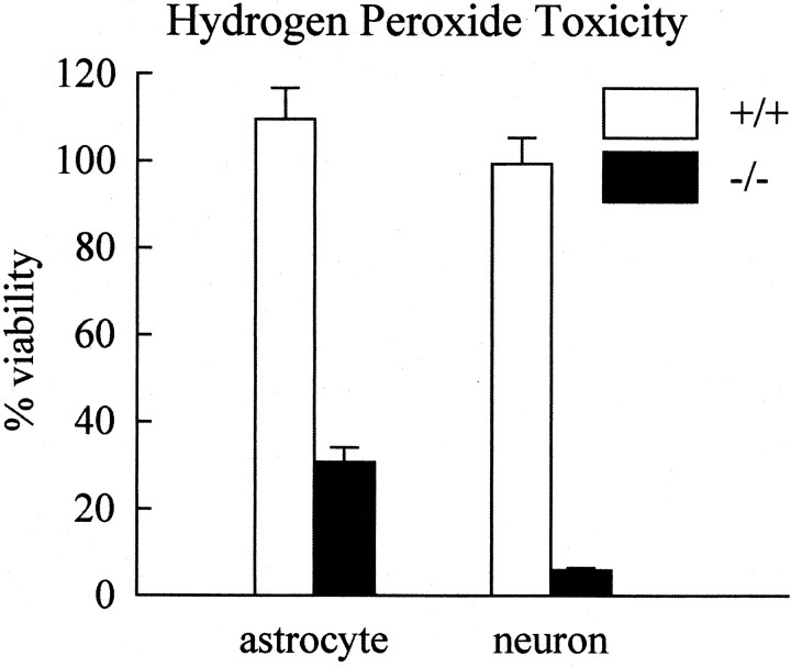
Ceruloplasmin is a ferroxidase that oxidizes toxic ferrous iron to its nontoxic ferric form. We have previously reported that a glycosylphosphatidylinositol-anchored form of ceruloplasmin is expressed in the mammalian CNS. To better understand the role of ceruloplasmin in iron homeostasis in the CNS, we generated a ceruloplasmin gene-deficient (Cp(-/-)) mouse. Adult Cp(-/-) mice showed increased iron deposition in several regions of the CNS such as the cerebellum and brainstem. Increased lipid peroxidation was also seen in some CNS regions. Cerebellar cells from neonatal Cp(-/-) mice were also more susceptible to oxidative stress in vitro. Cp(-/-) mice showed deficits in motor coordination that were associated with a loss of brainstem dopaminergic neurons. These results indicate that ceruloplasmin plays an important role in maintaining iron homeostasis in the CNS and in protecting the CNS from iron-mediated free radical injury. Therefore, the antioxidant effects of ceruloplasmin could have important implications for various neurodegenerative diseases such as Parkinson's disease and Alzheimer's disease in which iron deposition is known to occur.


Figures

Fig. 1.
Fig. 1.
Generation ofCp−/− mice. A, The targeting vector was generated by replacing the 3 kbBglII–NheI fragment containing the first exon of the Cp gene with a neo cassette. Homologous recombination of the targeting vector in ES cells leads to the generation of a 6.5 kb EcoRI fragment compared with a 11 kb wild-type fragment detected using a 1.5 kb probe upstream of the targeting vector (probe). BI, BglI;BII, BglII; EI,EcoRI; NI, NheI;PI, PstI; XI,XhoI. B, Southern blot ofEcoRI-digested genomic DNA from F2 animals using the probe shown in A. C, Western blot of serum using a rabbit anti-Cp polyclonal antibody. BothCp+/+ andCp+/− mice contain large amounts of Cp in the serum, whereasCp−/− mice lack Cp.D, Histological sections of the livers of 16-month-oldCp+/+ andCp−/− mice stained for iron using Perl's stain. The sections were counterstained with neutral red. The liver from a Cp+/+ mouse (+/+) does not show staining for iron. Only theneutral red counterstaining is visible. In contrast, the liver from a Cp−/−(−/−) mouse shows strong labeling for iron, which appears as blue staining.
Fig. 2.
Fig. 2.
Iron content and lipid peroxidation in CNS.A, The total non-heme iron content of different brain regions from Cp+/+ and Cp−/− mice. Values are shown as the mean ± SE. n = 4 for both groups of mice (*p ≤ 0.05; Student's t test).B, Levels of lipid peroxidation as assessed by measuring the levels of 2-thiobarbituric acid reactive substances in tissue homogenates of different brain regions from wild-type and knock-out mice are displayed. Values are shown as the mean ± SE.n = 4 for both groups of mice (*p ≤ 0.05; Student's ttest).
Fig. 3.
Fig. 3.
Iron histochemistry and Neurodegeneration.A, B, Iron histochemistry of the cerebellum of Cp+/+(A) and Cp−/−mice (B). Iron deposits, which appearbrown in color (arrows), are seen in the granule cell layer in Cp−/− mice (B). The inset in Bshows these iron deposits at higher magnification. Scale bar, 100 μm.C, D, TH+ dopaminergic neurons in the substantia nigra ofCp+/+ (C) andCp−/− mice (D). There are fewer TH+neurons in the null mice (D), and those present appear to be undergoing degeneration. Scale bars, 100 μm.
Fig. 4.
Fig. 4.
Retinal degeneration. A,B, Retina of 18-month-oldCp+/+ (A) andCp−/− mice (B) in Epon-embedded sections stained with toluidine blue. Neurodegenerative changes are seen in the inner nuclear layer (INL) of Cp−/−mice (B). ONL, Outer nuclear layer; GCL, ganglion cell layer. C,D, Higher magnification of the inner nuclear layer ofCp+/+ andCp−/− mice, respectively. Neurons in this layer in wild-type mice are large rounded cells with a prominent nucleolus (C). In contrast, inCp−/− mice many of the cells in this layer show condensed chromatin and dark cytoplasmin (D, arrows). Cells with small, irregularly shaped nuclei that are likely to be macrophages are also present in Cp−/− mice (D). Scale bars (shown in B forA, B): 50 μm; (shown inD for C, D): 20 μm.
Fig. 5.
Fig. 5.
Susceptibility of cerebellar cultures to H2O2 toxicity. A, Cultures of dissociated cerebellum were treated with H2O2, and their viability was assessed using MTT assay. Viability is expressed relative to untreated (0 μm H2O2) control cultures and is shown as the mean ± SE. Results are from three separate experiments. There is a significant decrease in cell viability in cultures of cerebellar cells fromCp−/− mice as compared with those from Cp+/+ mice treated with 50 μm H2O2 (*p≤ 0.05; Student's tTest).B, Cultures of dissociated cerebellum were treated with H2O2 in the presence of 125 μmDFO mesylate. Viability was assessed as above and is the result of three separate experiments. DFO treatment eliminated the decrease in viability of the knockout cultures mediated by the 50 μmH2O2 concentration. DFO partially prevented the decrease in viability caused by the 250 μmH2O2 concentration, and it rescued the cultures from Cp+/+ mice to a significantly greater extent (*p ≤ 0.05; Student'st test).
Fig. 6.
Fig. 6.
Counts of astrocyte and neuronal populations in cerebellar cultures from Cp+/+ andCp−/− mice treated with 50 μm H2O2 indicate that although the viability of both cell types is reduced in cultures ofCp−/− mice, the loss of neurons is more drastic than that of astrocytes. Results are presented as percentage viability compared with untreated cultures.
Fig. 7.
Fig. 7.
Tests of motor coordination. Motor coordination ofCp+/+ andCp−/− mice was assessed using the rotary rod assay. Mice were placed on a rotating rod, and the time, in seconds, that the mice remained on the rod without falling off, up to a maximum of 180 sec, was recorded. Mice of 16 months of age were used. The results are the shown as the mean ± SE; n= 4 for each group. A significant impairment in the ability ofCp−/− mice to remain on the rotary rod compared with Cp+/+ mice is observed (p ≤ 0.05; Student'st test).

References

    1. Andrews NC. The iron transporter DMT1. Int J Biochem Cell Biol. 1999;31:991–994. - PubMed
    1. Boll M-C, Sotelo J, Otero E, Alcaraz-Zubeldia M, Rios C. Reduced ferroxidase activity in the cerebrospinal fluid from patients with Parkinson's disease. Neurosci Lett. 1999;265:155–158. - PubMed
    1. Borg DC, Schaich KM. Prooxidant action of desferrioxamine: Fenton-like production of hydroxyl radicals by reduced ferrioxamine. J Free Radic Biol Med. 1986;2:237–243. - PubMed
    1. Bowie AG, Moynagh PN, O'Neill LAJ. Lipid peroxidation is involved in the activation of NF-kappaB by tumor necrosis factor but not interleukin-1 in the human endothelial cell line ECV304. Lack of involvement of H2O2 in NF-kappaB activation by either cytokine in both primary and transformed endothelial cells. J Biol Chem. 1997;272:25941–25950. - PubMed
    1. Choi SY, Kwon HY, Kwon OB, Eum WS, Kang JH. Fragmentation of human ceruloplasmin induced by hydrogen peroxide. Biochimie. 2000;82:175–180. - PubMed

Publication types

MeSH terms