. 2002 Aug 1;22(15):6623-30.
doi: 10.1523/JNEUROSCI.22-15-06623.2002.

Remyelination of the rat spinal cord by transplantation of identified bone marrow stromal cells

Affiliations

Remyelination of the rat spinal cord by transplantation of identified bone marrow stromal cells

Yukinori Akiyama et al. J Neurosci. .

Abstract

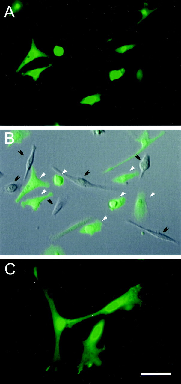
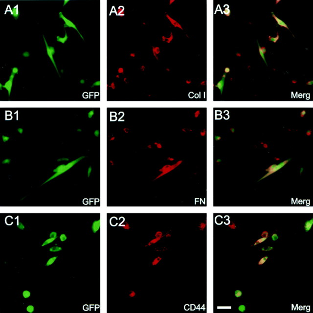
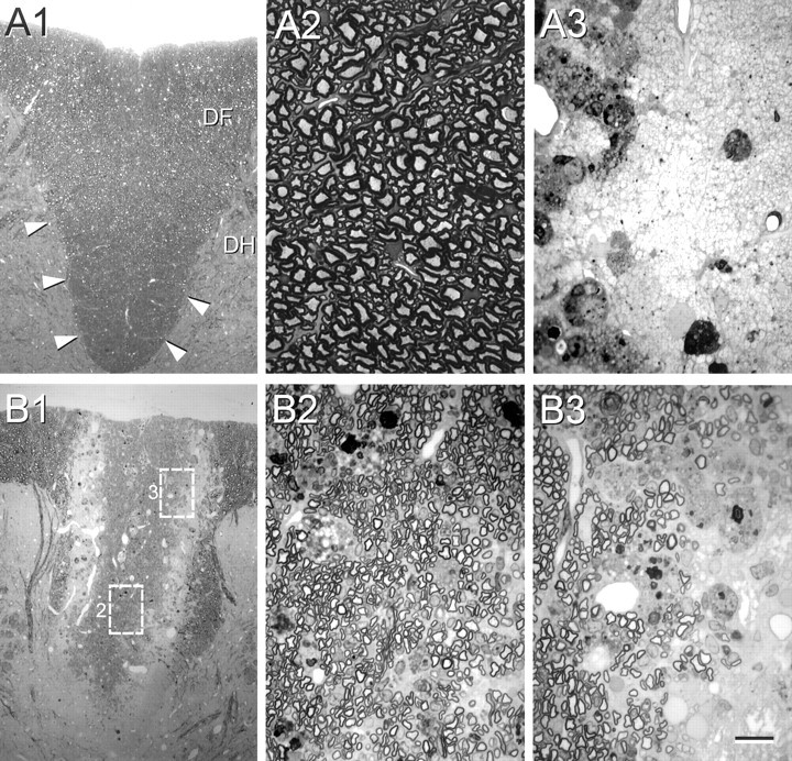
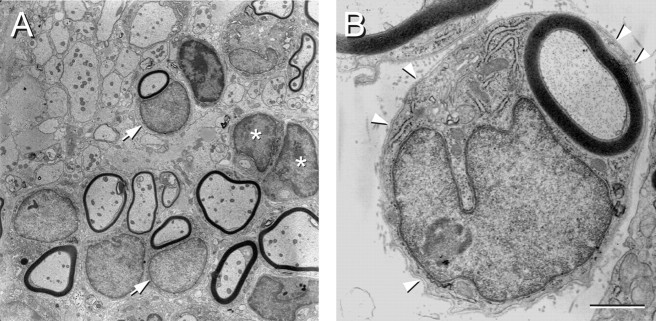
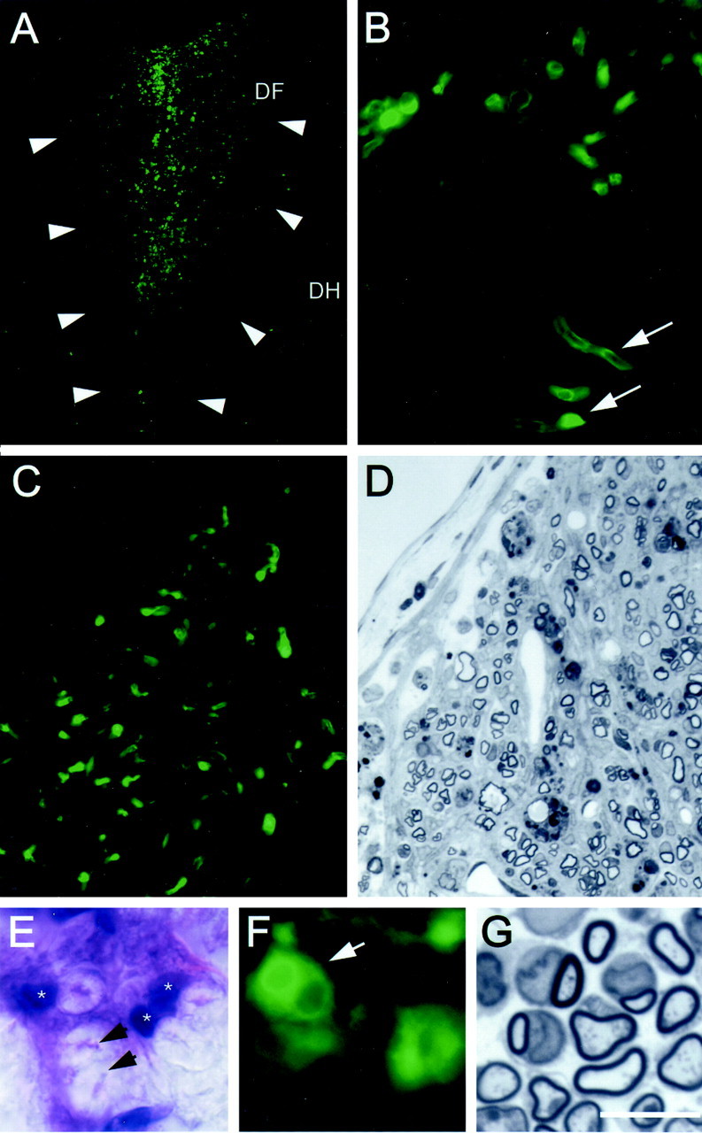
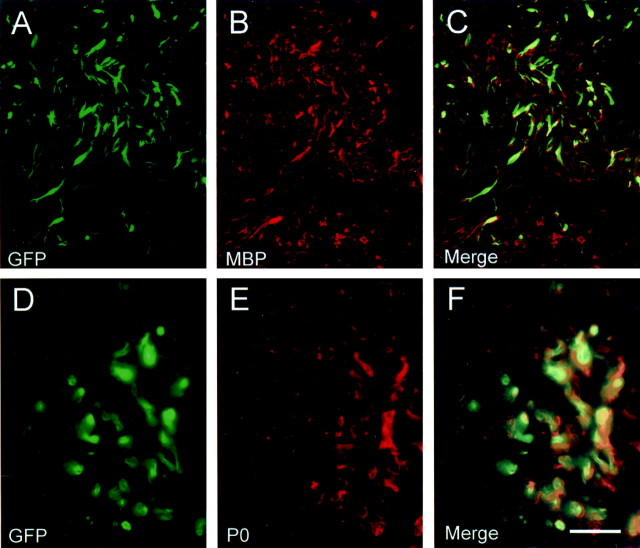
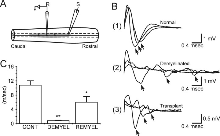
Bone marrow contains a population of stem-like cells that can differentiate into neurons or glia. Stromal cells from green fluorescent protein (GFP)-expressing mice were isolated by initial separation on a density gradient and then cultured as adherent cells on plastic that proliferated in culture to confluency with a typical flattened elongative morphology. The large majority of the isolated stromal cells were GFP expressing and immunopositive for collagen type I, fibronectin, and CD44. Transplantation of these cells by direct microinjection into the demyelinated spinal cord of the immunosuppressed rat resulted in remyelination. The remyelinated axons showed characteristics of both central and peripheral myelination as observed by electron microscopy; conduction velocity of the axons was improved. GFP-positive cells and myelin profiles were observed in the remyelinated spinal cord region, indicating that the donor-isolated stromal cells were responsible for the formation of the new myelin. The GFP-positive cells were colocalized with myelin basic protein-positive and P0-positive cellular elements. These findings indicate that cells contained within the stromal cell fraction of the mononuclear cell layer of bone marrow can form functional myelin during transplantation into demyelinated spinal cord.


Figures

Fig. 1.
Fig. 1.
GFP-expressing mouse bone marrow stromal cells cultured for 14 d. A, A field of GFP-expressing cells and superimposition on a DIC image of the same field (B). The cells vary in morphology from flattened and fusiform to rounded. White arrows indicate positive and black arrows indicate negative fluorescent cells. A higher-power image (C) shows a collection of GFP-expressing cells, which are flattened and process bearing. Scale bar: A, B, 50 μm; C, 30 μm.
Fig. 2.
Fig. 2.
Immunocytochemical characterization of cultured GFP-expressing mouse bone marrow stromal cells. Green fluorescence (A1, B1, C1) shows GFP-expressing bone marrow stromal cells from three different cultures (14 d in culture). Red fluorescence (A2,B2, C2) shows immunoreactivity to collagen type I (Col I), fibronectin (FN), and CD44, respectively. A3,B3, and C3 are coregistered images from their respective panels in 1 and 2. The large majority of GFP fluorescent cells were positive for collagen type I, fibronectin, and CD44. Scale bar, 10 μm.
Fig. 3.
Fig. 3.
Morphology of control, demyelinated, and remyelinated dorsal funiculus. Normal dorsal funiculus (area outlined by arrowheads in A1) of the T-11 spinal cord is shown at low (A1) and high (A2) power. A3, High-power field of demyelinated axons in control lesion with injection of control vehicle without cells. B1, A low-power micrograph of the dorsal funiculus lesion zone 3 weeks after GFP mouse bone marrow stromal cell transplantation. The high-power fields in B2 andB3 were obtained from the boxed areas inB1 marked 2 and 3, respectively. Note that nearly all of the axons in the central lesion zone in B2 are remyelinated, but that demyelinated axons are present at the lateral edge of the lesion (B3).DF, Dorsal funiculus; DH, dorsal horn. Scale bar: A1, B1, 100 μm;A2, A3, B2,B3, 15 μm. All images were obtained with Nikon Eclipse 800 epifluorescent microscope.
Fig. 4.
Fig. 4.
Electron micrographs of remyelinated axons in the dorsal funiculus of the spinal cord after bone marrow stromal cell transplantation. A, A field of axons showing some with myelin associated with a large cytoplasmic and nuclear surround, demyelinated axons, and axons surrounded by cytoplasmic regions but not yet forming myelin. Some of the myelinated axons did have the large nuclear and cytoplasmic surrounds and others without. B, A higher-power electron micrograph illustrating a remyelinated axon characteristic of a peripheral myelination pattern. Note the large cytoplasmic and nuclear domains and the basement membrane (arrowheads) surrounding the entire axon and cellular complex. Scale bar: A, 5 μm; B, 1 μm.
Fig. 5.
Fig. 5.
Identification of donor bone marrow cellsin vivo. A, Dorsal funiculus (DF) of coronally cut spinal cord from a rat that was injected with bone marrow stromal cells from GFP-expressing mice 3 weeks after transplantation. Arrows indicate the lateral margins of the dorsal funiculus. Numerous GFP-positive cells are observed in the remyelinated region. DH, Dorsal horn. B, Higher-power image of same field showing profiles reminiscent of myelinated axons. C and D are from frozen and plastic-embedded sections, respectively, from the same animal showing colocalization of GFP fluorescence and more clearly defined myelination in the plastic section. E andF show an H–E-stained frozen section and a fluorescent unstained image with GFP fluorescence at the same high power. Note that, in the frozen H–E section, the axon cylinder is collapsed (arrows) and the myelin is “puffy,” as is typical with this staining technique. G shows a comparable semithin plastic section from the same animal showing myelinated axons. The myelin is better preserved and the tissue more shrunken form dehydration protocols required for plastic embedding. Scale bar:A, 250 μm; B, 50 μm;C, D, 40 μm; E,F, 12 μm; G, 10 μm.
Fig. 6.
Fig. 6.
Colocalization of MPB and P0 with transplanted GFP-expressing stromal cells. Cryosections from spinal cord blocks adjacent to plastic-embedded blocks in which remyelination was confirmed showed colocalization of MBP (A–C) and P0 (D–F) with GFP-expressing elements. Images inA–C were obtained on a confocal microscope, andD–F were obtained on a conventional epifluorescence microscope. Scale bar: A–C, 70 μm;D–F, 30 μm.
Fig. 7.
Fig. 7.
Conduction velocity measurements of the remyelinated axons. A, Schematic indicating sites of stimulation (S) and recording (R) within the dorsal funiculus.B, Compound action potentials recorded at 1.0 mm increments longitudinally along the dorsal columns in normal (1), demyelinated (2), and after stromal cell transplantation (3). The arrows in B indicate the peak negativities of three primary negativities. C, Histograms of conduction velocity (error bars indicate SEM) of dorsal column axons obtained from normal (CONT), demyelinated (DEMYEL), and remyelinated (REMYEL) axons. At 36°C, conduction velocity of the early negativity in controls, demyelinated, and bone marrow-delivered groups were 10.76 ± 1.24 m/sec (n = 12), 0.89 ± 0.11 m/sec (n = 12), and 6.03 ± 1.62 m/sec (n = 9), respectively. *p < 0.01, demyelinated versus transplanted; **p < 0.001, control versus demyelinated.

References

    1. Akiyama Y, Honmou O, Kato T, Uede T, Hashi K, Kocsis JD. Transplantation of clonal neural precursor cells derived from adult human brain establishes functional peripheral myelin in the rat spinal cord. Exp Neurol. 2001;167:27–39. - PubMed
    1. Akiyama Y, Radtke C, Honmou O, Kocsis JD. Remyelination of the spinal cord axons by intravenous delivery of bone marrow cells. Glia. 2002;39:227–234. - PMC - PubMed
    1. Ashton BA, Allen TD, Howlett CR, Eaglesom CC, Hattori A, Owen M. Formation of bone and cartilage by marrow stromal cells in diffusion chambers in vivo. Clin Orthop. 1980;151:294–307. - PubMed
    1. Azizi SA, Stokes D, Augelli BJ, DiGirolamo C, Prockop DJ. Engraftment and migration of human bone marrow stromal cells implanted in the brains of albino rats—similarities to astrocyte grafts. Proc Natl Acad Sci USA. 1998;95:3908–3913. - PMC - PubMed
    1. Berthold C-H. Morphology of normal peripheral axons. In: Waxman SG, editor. Physiology and pathobiology of axons. Raven; New York: 1978. pp. 3–64.

Publication types

MeSH terms