. 2002 Aug 1;22(15):6631-8.
doi: 10.1523/JNEUROSCI.22-15-06631.2002.

Electrical stimulation promotes motoneuron regeneration without increasing its speed or conditioning the neuron

Affiliations

Electrical stimulation promotes motoneuron regeneration without increasing its speed or conditioning the neuron

Thomas M Brushart et al. J Neurosci. .

Abstract

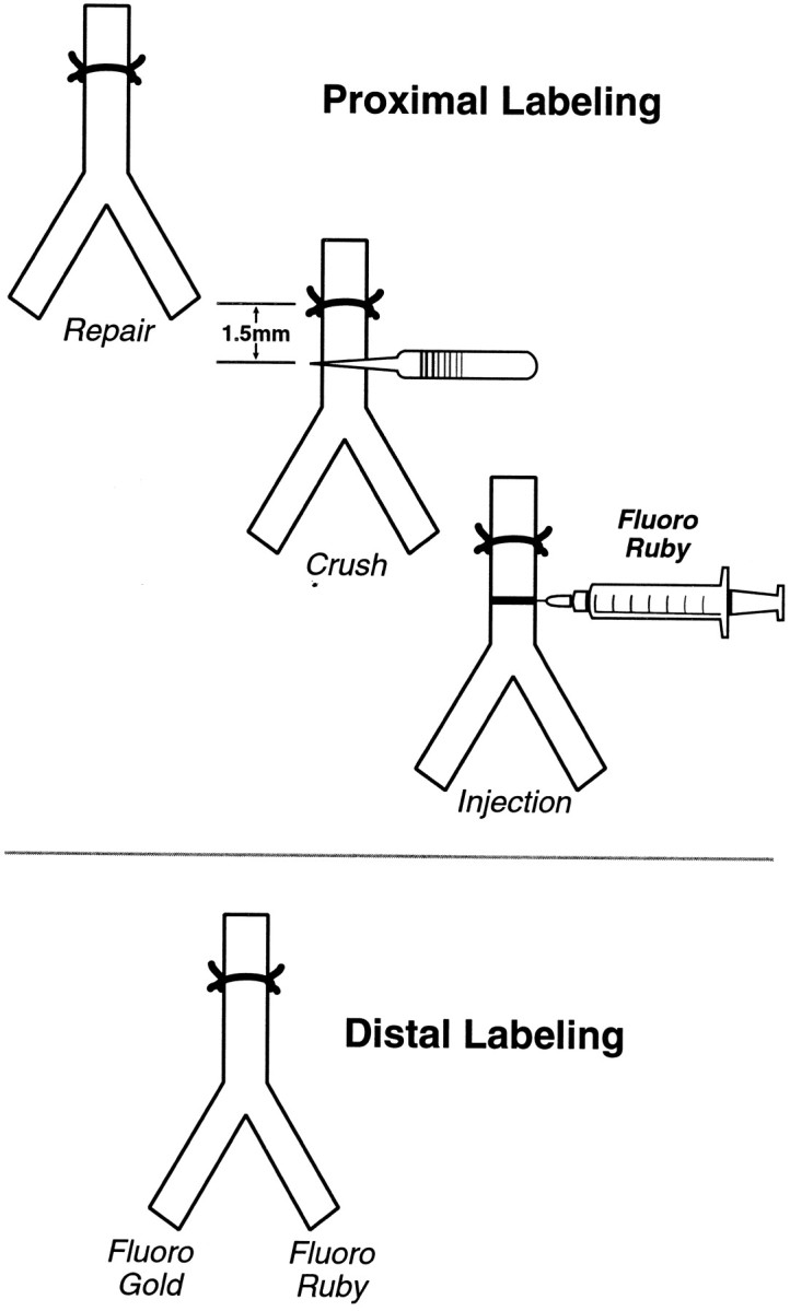
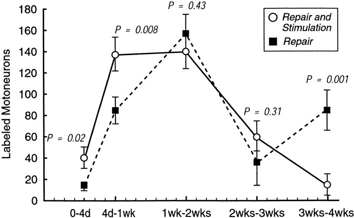
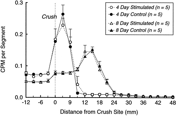
Motoneurons reinnervate the distal stump at variable rates after peripheral nerve transection and suture. In the rat femoral nerve model, reinnervation is already substantial 3 weeks after repair, but is not completed for an additional 7 weeks. However, this "staggered regeneration" can be temporally compressed by application of 20 Hz electrical stimulation to the nerve for 1 hr. The present experiments explore two possible mechanisms for this stimulation effect: (1) synchronization of distal stump reinnervation and (2) enhancement of regeneration speed. The first possibility was investigated by labeling all motoneurons that have crossed the repair at intervals from 4 d to 4 weeks after rat femoral nerve transection and suture. Although many axons did not cross until 3-4 weeks after routine repair, stimulation significantly increased the number crossing at 4 and 7 d, with only a few crossing after 2 weeks. Regeneration speed was studied by radioisotope labeling of transported proteins and by anterograde labeling of regenerating axons, and was not altered by stimulation. Attempts to condition the neuron by stimulating the femoral nerve 1 week before injury were also without effect. Electrical stimulation thus promotes the onset of motor axon regeneration without increasing its speed. This finding suggests a combined approach to improving the outcome of nerve repair, beginning with stimulation to recruit all motoneurons across the repair, followed by other treatments to speed and prolong axonal elongation.


Figures

Fig. 1.
Fig. 1.
Proximal and distal labeling. Proximal labeling identifies motor axons as soon as possible after they have entered the distal stump, but it does not differentiate between cutaneous and muscle pathways. Distal labeling is performed after axons have been segregated into cutaneous and muscle branches, revealing their destination but not their early behavior.
Fig. 2.
Fig. 2.
Cumulative distal stump reinnervation. The mean total numbers of motoneurons that have crossed the repair site with and without stimulation are plotted as a function of time after repair. Significantly more motoneurons have crossed in the stimulated nerves at all times during the first 3 weeks.
Fig. 3.
Fig. 3.
New crossing events. Each value was obtained by subtracting the mean number of motoneurons labeled at the beginning of an interval from the mean number labeled at the end of the interval for both stimulated and nonstimulated groups. The means at the 0–4 d interval are essentially the 4 d means, because a mean of only two motoneurons were labeled at the time of repair. The means at 4 d to 1 week in this figure represent the total number of motoneurons labeled at 1 week (Fig. 2) less the 4 d values, and so forth for each interval. Stimulation causes a rapid increase in crossing events to a peak between 1 and 2 weeks, with a more gradual decline. In contrast, the control curve is biphasic with a more gradual rise to an initial peak at 2 weeks, followed by rapid decline and then a second peak at 4 weeks. Electrical stimulation thus recruits many motoneurons to regenerate across the suture line and penetrate the distal stump earlier than they normally would.
Fig. 4.
Fig. 4.
Regeneration speed. Sciatic nerve crush with or without stimulation was followed in 3 or 7 d by injection of [35S]methionine into the parent motoneuron pool, and counts were obtained from peripheral nerve at 4 and 8 d, respectively. Counts per minute are plotted as a function of distance from the crush site. Each curve represents the mean of five nerves, normalized to facilitate comparison. Electrical stimulation had no effect on regeneration speed at either 4 or 8 d after crush.
Fig. 5.
Fig. 5.
Confocal microscopy of labeled motoneurons and axons. A–C represent femoral motoneurons retrogradely labeled with Fluoro Ruby (Scale bar, 100 μm). A,Normal femoral motoneuron pool; B, femoral motoneuron pool 1 week after nerve repair without stimulation. Motoneurons were labeled by crushing the nerve 1.5 mm distal to the repair and injecting the crush site with Fluoro Ruby (proximal labeling technique, Fig. 1). This technique labels axons as soon as they have entered the distal stump. C, Femoral motoneuron pool 1 week after nerve repair with stimulation, demonstrating an increase in the number of motoneurons that have crossed the repair site. D–F,Peripheral nerve double-labeled by anterograde transport of Neurobiotin (red) and antibodies to laminin (green) (Scale bar, 10 μm). D,Normal nerve 8 mm distal to the injection site, demonstrating nearly complete labeling of the nerve. E, Longitudinal section of normal nerve. The myelin sheath remains unlabeled. F,Axon reinnervating the distal stump. The axon is closely opposed to the basement membrane and is clearly identifiable by its growth cone. The Schwann cell tube below the axon contains fragmented diffusion artifact, which is confusing when viewed in cross-section but easily identified by its contour on longitudinal section. G,Profusely branched axon with multiple growth cones crossing the repair site of a stimulated animal. The axon is labeled with Neurobiotin only (Scale bar, 50 μm).
Fig. 6.
Fig. 6.
Conditioning trials–distal labeling. The femoral nerve was exposed bilaterally and stimulated unilaterally in 11 rats. One week later, both femoral nerves were transected and repaired. After allowing 3 weeks for regeneration, results were evaluated with distal labeling. For both groups of 11 nerves, the black barrepresents the mean number of motoneurons projecting correctly to the muscle branch, the white bar represents the mean number projecting incorrectly to the cutaneous branch, and the striped bar represents the mean number projecting to both branches (double-labeled). Stimulation 1 week before repair had no effect on the outcome of regeneration.
Fig. 7.
Fig. 7.
Conditioning trials–radiotracer. Sciatic nerve stimulation was followed in 1 week by crush. Label was injected 7 d after crush, and specimens were counted 1 d later. These animals do not differ significantly from the 8 d regeneration speed controls, and thus provide no evidence for a conditioning effect.

References

    1. Abercrombie M. Estimation of nuclear population from microtome sections. Anat Rec. 1946;94:239–247. - PubMed
    1. Al-Majed AA, Brushart TM, Gordon T. Electrical stimulation accelerates and increases expression of BDNF and trkB rnRNA in regenerating rat femoral motoneurons. Eur J Neurosci. 2000a;12:4381–4390. - PubMed
    1. Al-Majed AA, Neumann CM, Brushart TM, Gordon T. Brief electrical stimulation promotes the speed and accuracy of motor axonal regeneration. J Neurosci. 2000b;20:2602–2608. - PMC - PubMed
    1. Balkowiec A, Katz DM. Activity-dependent release of endogenous brain-derived neurotrophic factor from primary sensory neurons detected by ELISA in situ. J Neurosci. 2000;20:7417–7423. - PMC - PubMed
    1. Brushart TM. Preferential reinnervation of motor nerves by regenerating motor axons. J Neurosci. 1988;8:1026–1031. - PMC - PubMed

Publication types

LinkOut - more resources