. 2002 Aug 1;22(15):6639-49.
doi: 10.1523/JNEUROSCI.22-15-06639.2002.

The adult substantia nigra contains progenitor cells with neurogenic potential

Affiliations

The adult substantia nigra contains progenitor cells with neurogenic potential

D Chichung Lie et al. J Neurosci. .

Abstract

In Parkinson's disease, progressive loss of dopaminergic neurons in the substantia nigra pars compacta (SN) leads to debilitating motor dysfunction. One current therapy aims at exogenous cellular replacement of dopaminergic function by transplanting fetal midbrain cells into the striatum, the main projection area of the SN. However, results using this approach have shown variable success. It has been proposed that cellular replacement by endogenous stem/progenitor cells may be useful for therapeutic interventions in neurodegenerative diseases, including Parkinson's disease. Although it is widely accepted that progenitor cells are present in different areas of the adult CNS, it is unclear whether such cells reside in the adult SN and whether they have the potential to replace degenerating neurons. Here, we describe a population of actively dividing progenitor cells in the adult SN, which in situ give rise to new mature glial cells but not to neurons. However, after removal from the SN, these progenitor cells immediately have the potential to differentiate into neurons. Transplantation of freshly isolated SN progenitor cells into the adult hippocampus showed that these cells also have a neuronal potential under in vivo conditions. These results suggest that progenitor cells reside in the adult SN and can give rise to new neurons when exposed to appropriate environmental signals. This developmental potential of SN progenitor cells might be useful for future endogenous cell replacement strategies in Parkinson's disease.


Figures

Fig. 1.
Fig. 1.
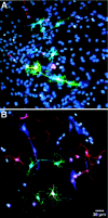
Proliferating cells are present in the adult SN. A, BrdU-labeled cells (green) are detected 2 hr after injection.B, At 3 d after injection, more BrdU-positive cells (green) are present in the SN. Cells are mostly found in doublets. A, B, TH in red.C, D, Proliferating cells are also detected by injection of a GFP retrovirus into the SN. D, Some of the infected cells express NG2 (red, colabeling with GFP inyellow). TH in red (C) or blue (D).
Fig. 2.
Fig. 2.
Phenotype of BrdU-positive cells in the SN after a 10 d BrdU pulse. A, One-half of the BrdU-positive cells (green) express the glial progenitor marker NG2 (blue). B, Some BrdU-positive cells (blue) that are not associated with blood vessels express the multipotent progenitor marker nestin (green). A, B, TH in shown inred.
Fig. 3.
Fig. 3.
Progenitor cells in the SN differentiate into glia 4 weeks after the final BrdU injection. A, Colocalization of a BrdU-positive nucleus (blue) with the oligodendrocyte marker APC (green).B, Colocalization of a BrdU-positive nucleus (blue) with the astrocyte marker S100β (green). C, Many BrdU-positive nuclei (blue) appear to be associated with dopaminergic neurons (red). Three-dimensional reconstruction in different planes (insets) revealed that these nuclei belong to satellite cells that are in close proximity to the dopaminergic neurons. S100β is shown in green.A–C, TH is shown in red.
Fig. 4.
Fig. 4.
Cultured SN progenitor cells give rise to all three neural lineages in vitro. β-tubulin III positive-neurons (A; green), GFAP-positive astrocytes (A; red), and RIP-positive oligodendrocytes (B; red) are observed after differentiation. A, B, Nuclei stained with DAPI (blue). C, BrdU was added 48 hr before differentiation. The nuclei of β-tubulin III-positive neurons (red) and GFAP-positive glia (blue) stain positive for BrdU (green), demonstrating that these cells are derived from a proliferating population.
Fig. 5.
Fig. 5.
Lineage potential of individual SN progenitor cells. Cells were infected with low-titer GFP retrovirus. Individual infected cells and their progeny were differentiated after a 7 d proliferation period in FGF8-supplemented media (A) or FGF2-supplemented media (B). After differentiation, some GFP-positive clones (green) produced β-tubulin III-positive neurons (red; seen as orange with GFP overlay) and GFAP-positive astrocytes (blue; seen asaqua with GFP overlay), demonstrating that the infected progenitor cell was multipotent. Nuclei stained with DAPI aredark blue.
Fig. 6.
Fig. 6.
In vivo proliferating SN progenitor cells have an intrinsic neuronal potential. Proliferating progenitor cells were labeled in vivo by injection of BrdU. SN progenitor cells were differentiated immediately after isolation. β-tubulin III-positive neurons (red) with BrdU-positive nuclei (green) were observed in all differentiation conditions tested: astrocyte feeder layer (A), forskolin-induced differentiation (B), and retinoic acid-induced differentiation (C).
Fig. 7.
Fig. 7.
In vivo neuronal differentiation potential of SN progenitor cells. A, BrdU-labeled SN progenitor cells (green) differentiate into NeuN (red)/β-tubulin III (blue)-positive neurons after transplantation to the hippocampus, demonstrating that SN progenitor cells can differentiate into neurons in vivo.B, In contrast, SN progenitor cells transplanted back to the SN do not differentiate into neurons, but display an NG2-positive glial progenitor phenotype (red), suggesting that the SN environment is not permissive for neuronal differentiation. BrdU is shown in green; TH is shown inblue.

References

    1. Altman J, Das GD. Autoradiographic and histological evidence of postnatal hippocampal neurogenesis in rats. J Comp Neurol. 1965;124:319–335. - PubMed
    1. Araque A, Carmignoto G, Haydon PG. Dynamic signaling between astrocytes and neurons. Annu Rev Physiol. 2001;63:795–813. - PubMed
    1. Bayer SA. Changes in the total number of dentate granule cells in juvenile and adult rats: a correlated volumetric and 3H-thymidine autoradiographic study. Exp Brain Res. 1982;46:315–323. - PubMed
    1. Bjorklund A, Lindvall O. Cell replacement therapies for central nervous system disorders. Nat Neurosci. 2000;3:537–544. - PubMed
    1. Cameron HA, McKay RD. Adult neurogenesis produces a large pool of new granule cells in the dentate gyrus. J Comp Neurol. 2001;435:406–417. - PubMed

Publication types

MeSH terms