. 2002 Aug 1;22(15):6724-31.
doi: 10.1523/JNEUROSCI.22-15-06724.2002.

Partial peripheral nerve injury promotes a selective loss of GABAergic inhibition in the superficial dorsal horn of the spinal cord

Affiliations

Partial peripheral nerve injury promotes a selective loss of GABAergic inhibition in the superficial dorsal horn of the spinal cord

Kimberly A Moore et al. J Neurosci. .

Abstract

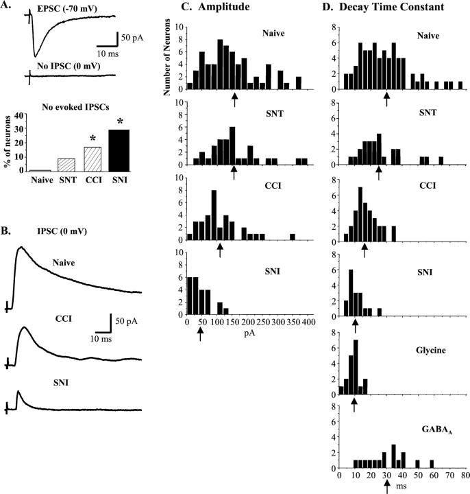
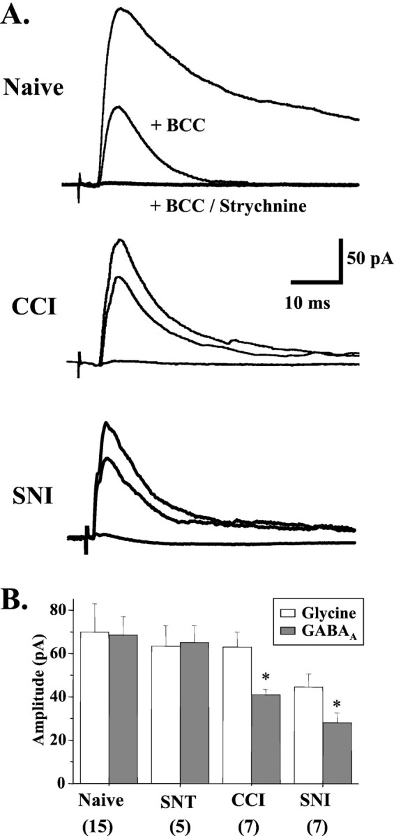
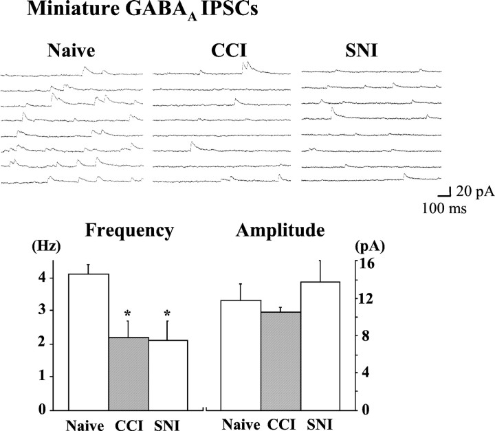
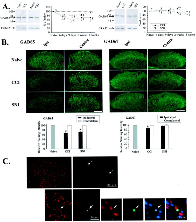
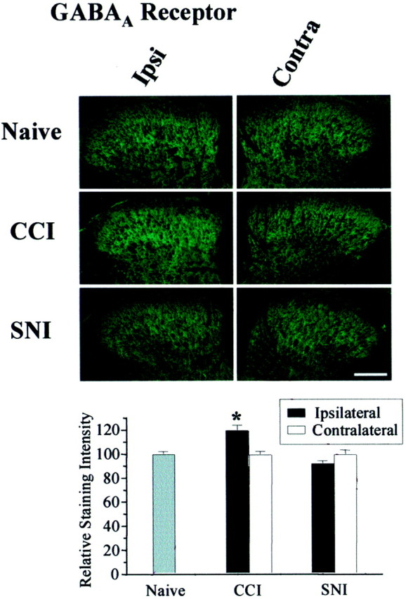
To clarify whether inhibitory transmission in the superficial dorsal horn of the spinal cord is reduced after peripheral nerve injury, we have studied synaptic transmission in lamina II neurons of an isolated adult rat spinal cord slice preparation after complete sciatic nerve transection (SNT), chronic constriction injury (CCI), or spared nerve injury (SNI). Fast excitatory transmission remains intact after all three types of nerve injury. In contrast, primary afferent-evoked IPSCs are substantially reduced in incidence, magnitude, and duration after the two partial nerve injuries, CCI and SNI, but not SNT. Pharmacologically isolated GABA(A) receptor-mediated IPSCs are decreased in the two partial nerve injury models compared with naive animals. An analysis of unitary IPSCs suggests that presynaptic GABA release is reduced after CCI and SNI. Partial nerve injury also decreases dorsal horn levels of the GABA synthesizing enzyme glutamic acid decarboxylase (GAD) 65 kDa ipsilateral to the injury and induces neuronal apoptosis, detected by terminal deoxynucleotidyl transferase-mediated biotinylated UTP nick end labeling staining in identified neurons. Both of these mechanisms could reduce presynaptic GABA levels and promote a functional loss of GABAergic transmission in the superficial dorsal horn.


Figures

Fig. 1.
Fig. 1.
Partial peripheral nerve injury (CCI or SNI) reduces primary afferent-evoked IPSCs in lamina II neurons.A, Representative neuron from a CCI animal with a monosynaptic Aδ-fiber-evoked EPSC but no evoked IPSC. After CCI or SNI, excitatory transmission remains intact, but the proportion of neurons with no evoked IPSC increases (Naive = 1/65; SNT = 3/32, p = 0.20;CCI = 7/41, p = 0.006;SNI = 9/32, p < 0.0001). *p < 0.05 compared with naive. B, Representative A-fiber-evoked IPSCs recorded in spinal cord slices from naive, CCI, and SNI rats. Note the reduction in amplitude and duration in CCI and SNI traces. C, Amplitude distributions for IPSCs recorded from naive rats and rats subjected to SNT, CCI, or SNI. In both partial nerve injury models, IPSC amplitudes were significantly decreased compared with the naive.Arrows indicate means. D, Frequency distributions of IPSC decay time constants (τ) from naive and nerve-injured rats, as well as pharmacologically isolated glycine (insensitive to 5–10 μm bicuculline;n = 18) and GABAA (bicuculline sensitive; n = 16) IPSCs.
Fig. 2.
Fig. 2.
Selective loss of GABAA IPSCs in CCI and SNI models. A, In naive neurons, the GABAA receptor antagonist bicuculline (5–10 μm) blocked a large fraction of the primary afferent-evoked IPSC (n = 14). However, after CCI (n = 13) or SNI (n = 7), bicuculline had little effect. Coapplication of bicuculline and strychnine (0.5 μm) reduced IPSCs to 4.3 ± 1.1 pA (n = 19). B, The amplitude of the GABAA component, but not the glycine component, of evoked IPSCs was significantly reduced after CCI (p= 0.014) or SNI (p = 0.002), but not SNT (p = 0.745). Numbers of neurons are indicated in parentheses. p< 0.05 compared with naive.
Fig. 3.
Fig. 3.
GABA transmission is reduced by a presynaptic mechanism after partial nerve injury. The frequency of miniature GABAA IPSCs (recorded in the presence of 0.5 μm TTX and 0.5 μm strychnine) was significantly decreased from 4.0 ± 0.3 Hz (n= 5) in naive slices to 2.2 ± 0.5 Hz (n = 8;p = 0.024) in CCI slices and 2.0 ± 0.6 Hz (n = 6; p = 0.020) in SNI slices. The amplitude of miniature GABAA IPSCs was unaffected by partial nerve injury (Naive = 11.7 ± 1.8 pA; CCI = 10.5 ± 0.6 pA;SNI = 13.8 ± 2.3 pA). p < 0.05 compared with naive.
Fig. 4.
Fig. 4.
Partial nerve injury reduces GAD65 levels and induces neuronal apoptosis. A, The 65 kDa, but not the 67 kDa, isoform of GAD was reduced in the lumbar (L4) dorsal horn ipsilateral to CCI or SNI when measured by Western blotting. Thegraph shows the time course of regulation. Data are normalized to ERK42 and expressed as a percentage of naive. *p < 0.05 compared with naive. B, Immunohistochemical analysis revealed diminished GAD65 levels in the ipsilateral dorsal horn (laminas I–IV) 2 weeks after CCI (n = 46 sections from 4 rats; p< 0.0001) and SNI (n = 31 sections from 6 rats;p < 0.0001). A small decrease in the levels of GAD67 was detectable after CCI (n = 52 sections from 4 rats; p = 0.002). Scale bar, 100 μm. *p < 0.05 compared with naive. C, TUNEL (green) and chromatin staining with bisbenzimide (blue), combined with immunostaining for the neuronal marker NeuN (red), revealed apoptotic cell death in the superficial dorsal horn of spinal cord segment L4 1 week after SNI. The top row shows a section through the ipsilateral dorsal horn with TUNEL-positive cells in laminas I and II (arrowheads). An example of neuronal apoptosis in lamina II is given below. Arrows point to a TUNEL-positive neuron with condensed chromatin structure, indicating nuclear pyknosis.
Fig. 5.
Fig. 5.
GABAA receptors remain after partial nerve injury. Immunohistochemical analysis for GABAAreceptor in the lumbar dorsal horn revealed a small upregulation after CCI (n = 27 sections from 3 rats;p = 0.031) but not SNI (n = 55 sections from 6 rats). Scale bar, 100 μm. Data are expressed as a percentage of naive. p < 0.05 compared with naive.

Similar articles

Cited by

References

    1. Alvarez FJ, Taylor-Blake B, Fyffe RE, De Blas AL, Light AR. Distribution of immunoreactivity for the β2 and β3 subunits of the GABAA receptor in the mammalian spinal cord. J Comp Neurol. 1996;365:392–412. - PubMed
    1. Baba H, Doubell TP, Woolf CJ. Peripheral inflammation facilitates Aβ fiber-mediated synaptic input to the substantia gelatinosa of the adult rat spinal cord. J Neurosci. 1999;19:859–867. - PMC - PubMed
    1. Bennett GJ, Xie YK. A peripheral mononeuropathy in rat that produces disorders of pain sensation like those seen in man. Pain. 1988;33:87–107. - PubMed
    1. Bennett GJ, Kajander KC, Saraha Y, Iadarola MJ, Sugimoto T. Neurochemical and anatomical changes in the dorsal horn of rats with an experimental peripheral neuropathy. In: Cervero F, Bennett GJ, Headley PM, editors. Processing of sensory information in the superficial dorsal horn of the spinal cord. Plenum; New York: 1989. pp. 463–471.
    1. Bhisitkul RB, Kocsis JD, Gordon TR, Waxman SG. Trophic influence of the distal nerve segment on GABAA receptor expression in axotomized adult sensory neurons. Exp Neurol. 1990;109:273–278. - PubMed

Publication types

MeSH terms