. 2002 Aug 20;99(17):11097-102.
doi: 10.1073/pnas.162138699. Epub 2002 Jul 31.

Closure of the Venus flytrap module of mGlu8 receptor and the activation process: Insights from mutations converting antagonists into agonists

Affiliations

Closure of the Venus flytrap module of mGlu8 receptor and the activation process: Insights from mutations converting antagonists into agonists

Anne-Sophie Bessis et al. Proc Natl Acad Sci U S A. .

Abstract

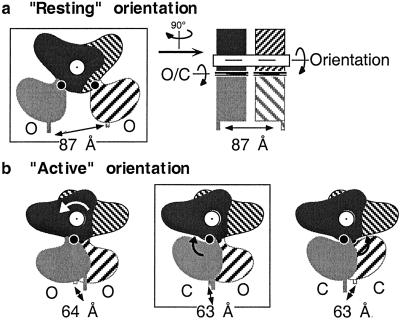
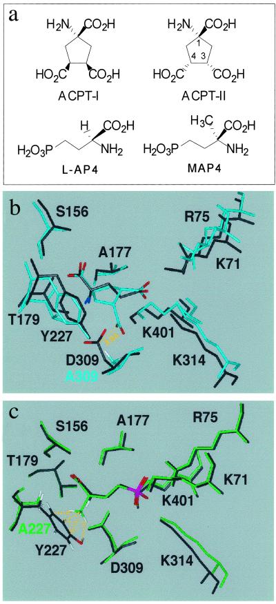
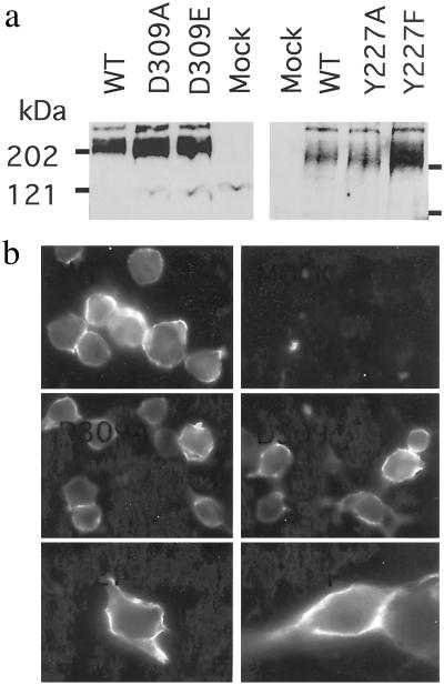
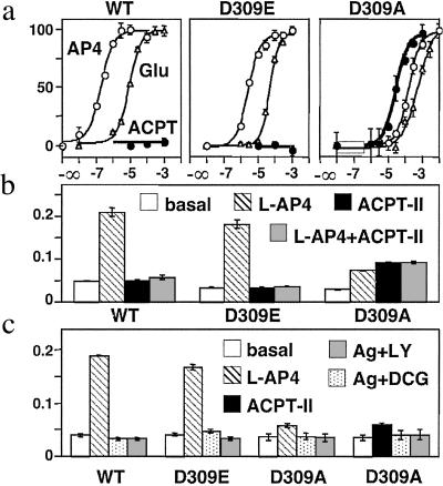
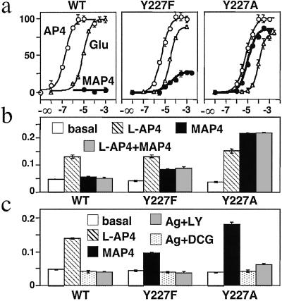
Ca2+, pheromones, sweet taste compounds, and the main neurotransmitters glutamate and gamma-aminobutyric acid activate G protein-coupled receptors (GPCRs) that constitute the GPCR family 3. These receptors are dimers, and each subunit has a large extracellular domain called a Venus flytrap module (VFTM), where agonists bind. This module is connected to a heptahelical domain that activates G proteins. Recently, the structure of the dimer of mGlu1 VFTMs revealed two important conformational changes resulting from glutamate binding. First, agonists can stabilize a closed state of at least one VFTM in the dimer. Second, the relative orientation of the two VFTMs in the dimer is different in the presence of glutamate, such that their C-terminal ends (which are connected to the G protein-activating heptahelical domain) become closer by more than 20 A. This latter change in orientation has been proposed to play a key role in receptor activation. To elucidate the respective role of VFTM closure and the change in orientation of the VFTMs in family 3 GPCR activation, we analyzed the mechanism of action of the mGlu8 receptor antagonists ACPT-II and MAP4. Molecular modeling studies suggest that these two compounds prevent the closure of the mGlu8 VFTM because of ionic and steric hindrance, respectively. We show here that the replacement of the residues responsible for these hindrances (Asp-309 and Tyr-227, respectively) by Ala allows ACPT-II or MAP4 to fully activate the receptors. These data are consistent with the requirement of the VFTM closure for family 3 GPCR activation.


Figures

Fig 1.
Fig 1.
Distance between the C-terminal ends of the VFTMs in the dimeric mGlu1 receptor extracellular domain depends on the relative orientation of the VFTMs. (a) Schemes depict the dimeric VFTMs in their “resting” orientation with both VFTMs in an open state, as observed in the crystal structure of the empty form (PDB ID code ) or the MCPG-bound form (PDB ID code ). One VFTM is in the front plane (gray), whereas the other is in the back plane (hatched). Lobe I is darker than lobe II. The right scheme corresponds to a view from the right side of the left scheme and highlights the main axis responsible for the change in conformation of this dimer. The axis for the rotation of one VFTM relative to the other is indicated in white, and the axis in each VFTM responsible for their closure is indicated in black. Models for the dimeric VFTMs in their resting orientation, with one or both modules in the closed state, reveal a similar 87-Å distance between their C-terminal ends (not shown). (b) Schemes represent the different possible conformations of the dimeric VFTMs in their “active” orientation, with both modules in an open state (Left) or one (Center) or both (Right) modules in the closed state. The latter two forms have been observed in crystals with bound glutamate (PDB ID code ) and glutamate and Gd3+ (PDB ID code ), respectively. The distances between the C-terminal ends of each module (α-carbon of Ile-512) in the resolved structures (boxed schemes) or 3D models are indicated. The models were obtained from the superposition of lobe I of the indicated VFTM conformers on lobe I of each subunit of the proposed resting (PDB ID code ) or active (PDB ID code ) dimers.
Fig 2.
Fig 2.
(a) Molecular structures of ligands. (b) 3D model of mGlu8-wt VFTM in its closed form with docked glutamate (black) and model of the D309A mutant with docked ACPT-II (cyan). C-α traces of both models have been superimposed. The distance between central carbon atom of C4-carboxylate of ACPT-II and that of Asp-309 is 3.0 Å, which results in repulsive ionic interaction in the wt. This repulsion is no longer present in mGlu8-D309A. (c) 3D model of mGlu8-wt in its closed form with docked L-AP4 and 3D model of Y227A mutant with docked MAP4. Similar superimposition between wt and Y227A models has been achieved as in b, showing a steric hindrance between the α-methyl group of MAP4 and Tyr-227, which is abolished in mGlu8-Y227A.
Fig 3.
Fig 3.
Expression and surface targeting of the wt, D309A, D309E, Y227F, and Y227A mGlu8 receptors. (a) Western blot analysis of the wild-type and mutant mGlu8a receptor showing the expected molecular mass for their dimeric form. (b) Cells expressing the indicated mGlu8a receptor epitope-tagged at their N-terminal extracellular end were labeled with the HA antibody. Cells were not permeabilized such that only the surface receptor could be detected.
Fig 4.
Fig 4.
ACPT-II is an agonist of mGlu8-D309A receptor. (a) Effect of increasing concentration of L-AP4 (○), glutamate (▵), or ACPT-II (•) on wt, D309E, and D309A mGlu8 receptors. Data are expressed as the percentage of the maximal effect of L-AP4. (b) ACPT-II is an antagonist at both wt and D309E mGlu8 receptors. IP production in cells expressing the indicated receptor under basal (open bars), L-AP4 (hatched bars), 1 mM ACPT-II (solid bars), or L-AP4 and 3 mM ACPT-II (shaded bars). L-AP4 concentrations used were 1.5 μM (wt), 10 μM (D309E), and 300 μM (D309A). Data are expressed as the IP production over the radioactivity remaining in the membranes. (c) Antagonist properties of DCG-IV and LY341495 (both at 1 mM) on wt, D309E, and D309A mGlu8 receptors. Values represent the IP production in cells expressing the indicated receptor under basal (open bars), L-AP4 (hatched bars), 30 μM ACPT-II (solid bars), the agonist plus DCG-IV (dotted bars), and the agonist plus LY341495 (shaded bars). L-AP4 concentrations used were 1.5 μM (wt), 10 μM (D309E), and 100 μM (D309A). Data are means ± SEM of triplicate determinations from typical experiments.
Fig 5.
Fig 5.
MAP4 is a partial agonist and a full agonist of mGlu8-Y227F and mGlu8-Y227A receptors, respectively. (a) Effect of increasing concentration of L-AP4 (○), glutamate (▵), or MAP4 (•) on wt, Y227F, and Y27A mGlu8 receptors. Data are expressed as the percentage of the maximal effect of L-AP4. (b) MAP4 is an antagonist at wt mGlu8 receptor and a partial agonist at mGlu8-Y227F receptor. IP production in cells expressing the indicated receptor under basal (open bars), L-AP4 (hatched bars), 3 mM MAP4 (solid bars), or L-AP4 and 3 mM MAP4 (shaded bars). L-AP4 concentration was 1.5 μM (wt) or 10 μM (Y227F and Y227A). Data are expressed as the IP production over the radioactivity remaining in the membranes. (c) Antagonist properties of DCG-IV (1 mM) and LY341495 (100 μM) on wt, Y227F, and Y227A mGlu8 receptors. Values represent the IP production in cells expressing the indicated receptor under basal (open bars), 1.5 μM L-AP4 (hatched bars), 100 μM MAP4 (solid bars), the agonist plus DCG-IV (dotted bars), and the agonist plus LY341495 (shaded bars). Data are means ± SEM of triplicate determinations from typical experiments.

References

    1. Bockaert J. & Pin, J.-P. (1999) EMBO J. 18, 1723-1729. - PMC - PubMed
    1. De Blasi A., Conn, P. J., Pin, J.-P. & Nicoletti, F. (2001) Trends Pharmacol. Sci. 22, 114-120. - PubMed
    1. Takahashi K., Tsuchida, K., Tanabe, Y., Masu, M. & Nakanishi, S. (1993) J. Biol. Chem. 268, 19341-19345. - PubMed
    1. O'Hara P. J., Sheppard, P. O., Thøgersen, H., Venezia, D., Haldeman, B. A., McGrane, V., Houamed, K. M., Thomsen, C., Gilbert, T. L. & Mulvihill, E. R. (1993) Neuron 11, 41-52. - PubMed
    1. Costantino G. & Pellicciari, R. (1996) J. Med. Chem. 39, 3998-4006. - PubMed

Publication types

MeSH terms

LinkOut - more resources