Comparative Study
. 2002 Aug 1;16(15):1890-905.
doi: 10.1101/gad.1004802.

Molecular mechanisms governing Pcdh-gamma gene expression: evidence for a multiple promoter and cis-alternative splicing model

Affiliations
Comparative Study

Molecular mechanisms governing Pcdh-gamma gene expression: evidence for a multiple promoter and cis-alternative splicing model

Xiaozhong Wang et al. Genes Dev. .

Abstract

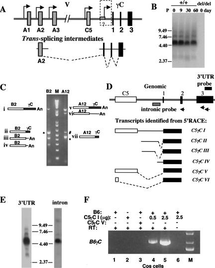
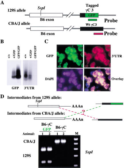
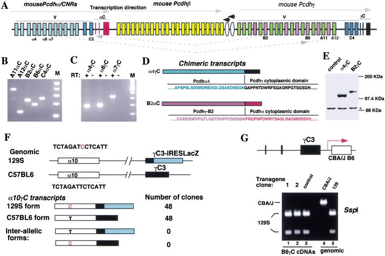
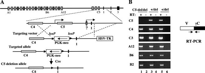
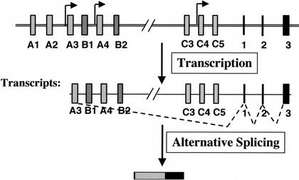
The genomic architecture of protocadherin (Pcdh) gene clusters is remarkably similar to that of the immunoglobulin and T cell receptor gene clusters, and can potentially provide significant molecular diversity. Pcdh genes are abundantly expressed in the central nervous system. These molecules are primary candidates for establishing specific neuronal connectivity. Despite the extensive analyses of the genomic structure of both human and mouse Pcdh gene clusters, the definitive molecular mechanisms that control Pcdh gene expression are still unknown. Four theories have been proposed, including (1) DNA recombination followed by cis-splicing, (2) single promoter and cis-alternative splicing, (3) multiple promoters and cis-alternative splicing, and (4) multiple promoters and trans-splicing. Using a combination of molecular and genetic analyses, we evaluated the four models at the Pcdh-gamma locus. Our analysis provides evidence that the transcription of individual Pcdh-gamma genes is under the control of a distinct but related promoter upstream of each Pcdh-gamma variable exon, and posttranscriptional processing of each Pcdh-gamma transcript is predominantly mediated through cis-alternative splicing.


Figures

Figure 1
Figure 1
Mouse Pcdh-γ locus and genetically modified alleles. Multiple modified Pcdh-γ alleles used in this study are shown in relation to the endogenous locus. Deletion allele, IRES-LacZ reporter allele, GFP fusion allele, WT-B6CBA/J transgene allele, Mut-B6CBA/J transgene allele, and C5 deletion allele.
Figure 2
Figure 2
A single Pcdh-γ allele expresses multiple variable exons. (A) DNA recombination model for Pcdh gene expression (Wu and Maniatis 1999). (B) Detection of multiple different Pcdh-γ genes using single-cell RT-PCR. Single neurons from Pcdhdel/GFP cerebellum (postnatal P5) were used to detect γC-GFP, A12-γC, A11-γC, and B2-γC.
Figure 3
Figure 3
No deletion of common intervening sequences upstream of the constant region. The majority of cells express Pcdh-γ in the cortex of adult mouse brain. (A) LacZ staining of a vibratome section (left) and a frozen section (right) from adult mouse brain (Pcdh+/LacZ); (B) Immunostaining of a frozen section of Pcdh+/GFP mouse brain with rabbit anti-GFP antibodies and mouse anti-SV2 antibody. (C) In situ hybridization of a frozen section from adult Pcdh+/GFP mouse brain using a fluorescein-labeled GFP riboprobe (in yellow) and stained with DAPI (in blue); (D) No deletion of common intervening sequences upstream from the constant region. Genomic DNAs were obtained from liver (L) and cortex (C ) of Pcdh+/del adult mice. Gene dosage comparison by Southern blot analysis using the three probes indicated. (E) Expression of multiple Pcdh-γ genes in heterozygous (Pcdh+/del) ES cells. Western blot analysis using anti-GFP antibodies detected Pcdh-γ-GFP fusion proteins in Pcdh+/GFP ES cells. RT-PCR analysis detected multiple spliced forms of Pcdh-γ cDNAs in Pcdh+/del ES cells.
Figure 4
Figure 4
Individual variable exon transcripts share no common 5′UTR sequence. (A) The single promoter and cis-alternative splicing model (Wu and Maniatis 1999). (B) 5′RACE analysis on two Pcdh-γ variable exon transcripts identified multiple transcription start sites immediately upstream of translation start ATG codon. These transcripts have a different 5′ UTR sequence.
Figure 5
Figure 5
Individual Pcdh-γ variable exons have their own promoters. (A) Identification of SNPs and RFLPs in variable exon B6 between 129S and CBA/J mouse strains. The relevant sequences from different mouse strains are shown as well as SspI RFLP analysis of genomic DNA from different mouse strains. (B) Targeting of a B6 transgene from the CBA/J strain into the Pcdh-γ locus in mouse ES cells (129S strain). The CBA/J B6 transgene consists of 2.5-kb 5′ flanking sequence, the coding exon and 2.1 kb of intronic sequence. This transgene is transcribed in the same orientation as the endogenous Pcdh-γ gene cluster. cDNA RFLP analysis revealed that the CBA/J-B6 exon was expressed at a level similar to the endogenous 129S-B6 exon (lanes 1,3). Wild-type ES cells serve as a control (lane 5). The RT (minus) control excludes the possibility of genomic DNA contamination.
Figure 6
Figure 6
A conserved DNA motif is required for the transcription of each variable exon. (A) Targeting of a mutant CBA/J B6 transgene. This mutant transgene includes a deletion of the 20-bp DNA motif present upstream of each variable exon. The mutant transgene was targeted to the same location as the wild-type (WT) transgene (Fig. 5A). (B) Both the WT transgene- and mutant transgene-targeted ES clones were differentiated into neurons in vitro. RT-PCR and RFLP analysis showed a significant decrease of the CBA/J B6 exon encoded by the mutant transgene. (C,D) Analysis of the relative ratio of the expressed CBA/J-B6 and 129S-B6 transcripts in the differentiated neurons (C) and undifferentiated ES cells (D).
Figure 7
Figure 7
The diversity of Pcdh-γ transcripts. (A) Multiple promoters and trans-splicing model (Wu and Maniatis 1999). (B) Northern blot analysis shows that a variety of Pcdh-γ transcripts are present at different stages of mouse brain development. (C) 3′RACE analysis of Pcdh-γ B2 and A12 transcripts identified both spliced (i, ii, v, and vii) and nonspliced transcripts (iii, iv, and vi). B2-ii and A12-vii are 3′RACE products generated by annealing of an oligo-dT primer in the A-rich region of constant exon 3. The # indicates a background band resulting from single oligo-dT primer amplification. The * indicates PCR fragments containing the chimeric transcripts B2αC or A12αC described in Fig. 9. (D) 5′RACE analysis identified a variety of constant exon-containing transcripts. The schematic drawing illustrates the genomic region containing the Pcdhγ C5 variable exon and three constant exons (not to scale) and alternatively spliced C5γC transcripts (I–VI). The primers used in the 5′RACE are depicted as arrows. (E) Northern blot analysis shows that the intron-containing Pcdh-γ transcripts are present in newborn mouse brain (intronic probe, right panel). (F) In vitro trans-splicing of the Pcdh-γ B6 variable exon with a C5γC intermediate. COS-7 cells were transfected with combinations of the indicated expression vectors. RT-PCR analysis of the RNA from transfected cells shows B6γC trans-spliced transcripts (lanes 4,5) between B6 and C5γC I. Note that trans-splicing did not occur in the cells coexpressing the B6 gene and the C5γC VI cDNA (lane 3).
Figure 8
Figure 8
In vivo assessment of trans-splicing between different Pcdh-γ alleles. (A) Allelic differences in both variable and constant exons. An SspI RFLP detected the allelic difference in the variable B6 exon. The second allelic difference in the constant region was introduced by tagging constant exon 3 with a GFP cDNA in the 129S allele. (B) Northern blot analysis using allele-specific probes shows that both Pcdh-γ alleles are active in transcription (comparing the hybridization signals between heterozygous and homozygous mice). (C) Both Pcdh-γ alleles are actively transcribed in single neurons. Shown is double-fluorescence-labelled in situ hybridization of frozen sections of adult mouse brain (Pcdh+/GFP), using allele-specific probes. (D) Trans-splicing between different alleles is not abundant. Two heterozygous mice were analyzed (1 and 2), each carrying both the targeted allele and CBA/J allele. RFLP analysis of two types of B6γC cDNAs demonstrated that the spliced B6γC transcripts predominantly originated from the same allele. B6γC-GFP cDNA was from the 129S allele; and B6γC cDNA was from the CBA/J allele. Interallelic B6γC transcripts were not abundant.
Figure 9
Figure 9
Pcdh-α/γ chimeric transcripts. (A) Schematic diagram of the mouse Pcdh gene clusters. Note that all three clusters are transcribed in the same direction, and two additional genes illustrated by ovals are transcribed in the opposite direction. (B) RT-PCR analysis identified chimeric transcripts between the Pcdh-γ variable exons A11, A12, B2, B6, and C4 and the Pcdh-α constant exons (αC) from mouse brain RNA. (C) RT-PCR analysis identified chimeric transcripts between the Pcdhα variable exons α4, α6, and α7 and the Pcdh-γ constant exons (γC). (D) The chimeric proteins (α4γC and B2αC) encoded by the chimeric transcripts. (E) Western blot analysis confirmed the expression of an in-frame α4γC protein in transfected cells. The * indicates a background band serving as the loading control. (F) Interallelic trans-splicing assay of α10γC chimeric transcripts (similar to Fig. 8). An SNP (red C on 129S strain α10 exon) and the targeted IRES-LacZ cassette define allelic differences in both α10 variable exon and γ constant exon 3. Sequence analysis of two different types of α10γC cDNAs showed that each originated from its original allele, suggesting that trans-splicing might have occurred on the same chromosome. (G) Trans-splicing is infrequent on the same chromosome within the Pcdh-γ cluster. The CBA/J-B6 transgene was targeted to the end of the Pcdh-γ locus and is transcribed in the same orientation as the endogenous locus. This mimics the configuration of the γ variable exon spliced to the α constant exons (in B). RT-PCR was used to amplify spliced B6γC cDNA from the undifferentiated ES cells using a specific primer for the constant exon. RFLP analysis of the RT-PCR products showed that splicing predominantly occurred from the endogenous 129S-B6 exon to the γ constant exons but not from the CBA/J-B6 exon (lanes 1 and 2, cf. lane 3 of control ES cells).
Figure 10
Figure 10
Cis-splicing in the absence of upstream sequence from constant exon 1. (A) Deletion of C5 and conserved upstream DNA sequences from constant exon 1. A 5-kb sequence containing the entire C5 gene and the conserved DNA sequences upstream of constant exon 1 was deleted by gene targeting in ES cells carrying only one Pcdh-γ allele. (B) RT-PCR analysis showed that individual variable exons (C3, C4, B2, B6, and A12) were efficiently spliced to γ constant exons in the absence of the 5-kb upstream sequence from constant exon 1.
Figure 11
Figure 11
Cell-specific expression of a subset of Pcdh genes: Differential promoter activation and cis-alternative splicing. In this model, individual cells express a distinct subset of protocadherins. The expression of the specific combination of protocadherins is achieved by differential promoter activation, followed by cis-alternative splicing.

References

    1. Agabian N. Trans splicing of nuclear pre-mRNAs. Cell. 1990;61:1157–1160. - PubMed
    1. Akopian AN, Okuse K, Souslova V, England S, Ogata N, Wood JN. Trans-splicing of a voltage-gated sodium channel is regulated by nerve growth factor. FEBS Lett. 1999;445:177–182. - PubMed
    1. Bain G, Kitchens D, Yao M, Huettner JE, Gottlieb DI. Embryonic stem cells express neuronal properties in vitro. Dev Biol. 1995;168:342–357. - PubMed
    1. Benson DL, Colman DR, Huntley GW. Molecules, maps and synapse specificity. Nat Rev Neurosci. 2001;2:899–909. - PubMed
    1. Bonen L. Trans-splicing of pre-mRNA in plants, animals, and protists. FASEB J. 1993;7:40–46. - PubMed

Publication types

MeSH terms

Associated data