Comparative Study
. 2002 Aug 1;16(15):1934-49.
doi: 10.1101/gad.984502.

Evidence of a critical architectural function for the RAG proteins in end processing, protection, and joining in V(D)J recombination

Affiliations
Comparative Study

Evidence of a critical architectural function for the RAG proteins in end processing, protection, and joining in V(D)J recombination

Chia-Lun Tsai et al. Genes Dev. .

Abstract

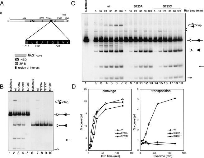
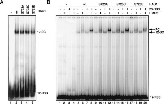
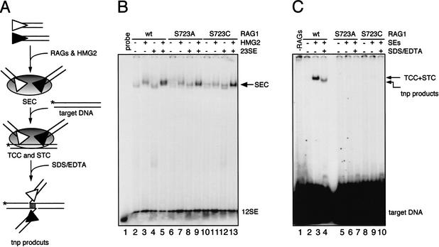
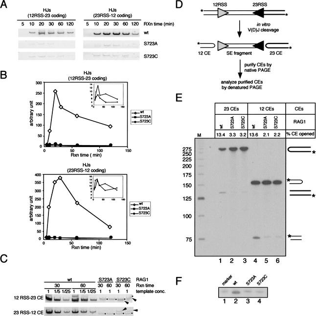
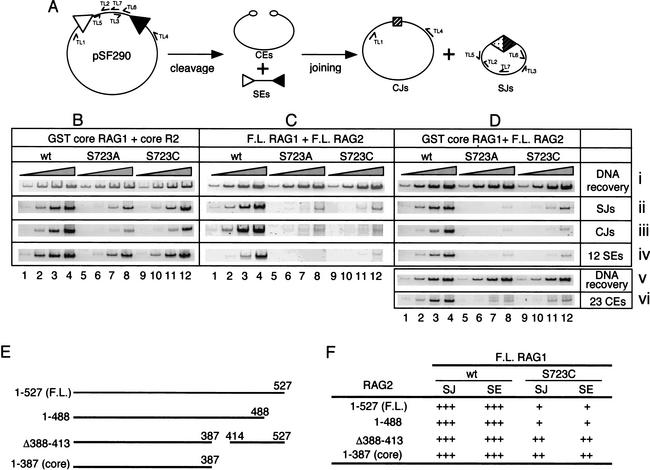
In addition to creating the DNA double strand breaks that initiate V(D)J recombination, the RAG proteins are thought to play a critical role in the joining phase of the reaction. One such role, suggested by in vitro studies, might be to ensure the structural integrity of postcleavage complexes, but the significance of such a function in vivo is unknown. We have identified RAG1 mutants that are proficient in DNA cleavage but defective in their ability to interact with coding ends after cleavage and in the capture of target DNA for transposition. As a result, these mutants exhibit severe defects in hybrid joint formation, hairpin coding end opening, and transposition in vitro, and in V(D)J recombination in vivo. Our results suggest that the RAG proteins have an architectural function in facilitating proper and efficient V(D)J joining, and a protective function in preventing degradation of broken ends prior to joining.


Figures

Figure 1
Figure 1
Coupled cleavage and intramolecular transposition. (A) Schematic diagram of the murine core RAG1 protein depicting three active site residues (D600, D708, and E962), the nonamer binding domain (NBD), the ZFB zinc finger domain, and the amino acid sequence surrounding S723. (B) Cleavage and transposition activities were assessed using a body-labeled cleavage substrate containing a 12-RSS and a 23-RSS (lanes 15) or a precleaved signal end fragment (lanes 610). Structures of the substrates and reaction products are indicated at the right, with the 12-RSS and 23-RSS depicted as open and filled triangles, respectively, and coding ends as open circles. The MBP-RAG1 protein used is indicated above each lane. tnp, intramolecular transposition product. (C) Kinetic analysis of coupled cleavage. The asterisks indicate background bands present in the substrate. (D) Quantitation of cleavage and transposition activities in (C). The cleavage activity was calculated as the ratio of the intensity of the signal end fragment (including both the cleavage product and transposition product) to the total radioactivity in each reaction. Transposition activity was calculated as the ratio of the intensity of the transposition product to the sum of the signal end fragment and the transposition product.
Figure 2
Figure 2
Formation of the 12-SC and the paired complex. (A) EMSA for detection of the 12-SC was performed using MBP-RAG1, GST-RAG2, and HMG2 proteins in 5 mM Ca++ with a 5′ end-labeled double stranded 12-RSS oligonucleotide substrate. Complexes were resolved on a native 4% polyacrylamide gel. (B) The assay for paired complex formation was performed with labeled 12-RSS in a buffer containing Ca++ in the presence or absence of unlabeled 23-RSS and HMG2 as indicated. Complexes were analyzed on a native 6% (80:1) polyacrylamide gel. The positions of the bands corresponding to the 12-SC as well as the paired complex are indicated at the right.
Figure 3
Figure 3
Formation of the SEC, the TCC, and intermolecular transposition products. (A) Schematic diagram of the complexes leading to transposition. The SEC is formed from a pair of signal ends, whereas in the TCC and STC, the signal ends are associated noncovalently or covalently, respectively, with target DNA (which can be revealed by denaturation with SDS/EDTA). (B) SEC formation reactions were carried out using 5′ end-labeled 12-signal end oligonucleotide in a reaction buffer containing 4 mM Mg++ in the presence or absence of HMG2 and unlabeled 23-signal end as indicated above the lanes. Reactions were analyzed on a native 6% (80:1) polyacrylamide gel. (C) TCC formation and intermolecular transposition reactions were carried out in a two-stage fashion. First, the SEC was formed as in (B) except that both the 12-signal end and 23-signal end were unlabeled. Formation of the TCC and STC were initiated by adding 5′ end-labeled oligonucleotide target DNA. Deproteinized transposition products were visualized by treating the reaction with SDS/EDTA prior to loading on a native 6% (80:1) polyacrylamide gel (lanes 4,7,11). Treatment with higher concentrations of EDTA (25 mM) together with 0.5% SDS yielded identical results (data not shown). The transposition product band seen in lane 4 is temperature-labile (55°C, 5′), as predicted from the structure shown at the bottom of panel A.
Figure 4
Figure 4
Stability of the postcleavage complex. (A) Schematic diagram of a biotin-labeled postcleavage complex bound to a streptavidin magnetic bead. (B) Large-scale coupled cleavage reactions were carried out with streptavidin bead-bound DNA substrate at 37°C for 1 h. Samples are indicated as: input, one-quarter of each reaction prior to purification; unbound, DNA in the reaction supernatant, not retained on the beads; wash, DNA in the supernatant of the second wash; elution, DNA eluted by treatment with SDS/EDTA; beads, DNA eluted by subsequent harsh treatment of the beads with proteinase K for 60 min at 55°C followed by phenol/chloroform extraction (this removes only ∼50% of the bound, biotinylated substrate from the beads; data not shown). wt, wild-type RAG1; SA, S723A RAG1; SC, S723C RAG1. Arrowheads indicate the signal end fragment resulting from coupled cleavage. Shown here is a representative gel from three independent experiments. (C) Quantitation of the stability of the postcleavage complex as measured by the ratio of intensity of the signal end fragment in the elution fraction to that in the input fraction (corrected for loading).
Figure 5
Figure 5
Postcleavage activities of the RAG proteins in vitro. (A) Hybrid joint formation. Products of the coupled cleavage reactions described in Fig. 1B were used as templates for PCR reactions to amplify hybrid joints. PCR products were resolved on 6% polyacrylamide gels and visualized by staining with cybergreen I. (B) Quantitation of hybrid joints in (A) by FluorImager (Molecular Dynamics). (C) Two time points (30 and 60 min) of the coupled cleavage reaction products were further analyzed by a semiquantitative PCR assay. The reactions containing the wild-type RAG1 protein were subjected to successive fivefold dilutions prior to PCR amplification, whereas the reactions containing the mutant RAG1 proteins were used as templates for PCR without dilution. Asterisks indicate nonspecific PCR products, and arrows indicate specific HJ products. (D) Schematic diagram showing the assay to detect hairpin opening by the RAG proteins in the context of coupled cleavage. Large-scale coupled cleavage reactions were carried out using a 5′ end-labeled PCR substrate, and the resulting coding ends were purified from a 4% native polyacrylamide gel. Purified coding ends were then analyzed on a 10% denaturing gel containing 40% (v/v) formamide, shown in (E). Unprocessed coding ends give rise to bands at 272 nt and 154 nt, whereas the processed coding ends yield products of ∼136 nt and 77 nt. The hairpin opening activity (% CE opened) is indicated above each lane and was determined by calculating the ratio of the processed coding end to the total coding end and multiplying by 100. (F) Purified 12-coding ends were further analyzed by comparing to a synthetic oligonucleotide identical in length and sequence to the top strand of the 12-coding end that has been nicked at the hairpin tip (lane 1). The processed coding ends in the wild-type RAG1 reaction migrated slightly faster than the marker and the processed coding ends from the reactions containing mutant RAG1 proteins (cf. lane 2 and lanes 1,3,4).
Figure 6
Figure 6
In vivo V(D)J recombination. (A) Schematic diagram of recombination of the pSF290 recombination substrate. Specific PCR primers used to amplify recombination intermediates and products are indicated. In vivo recombination assays were carried out with the combinations of (B) GST-core RAG1 and his-myc tagged core RAG2; (C) untagged full-length RAG1 and RAG2; and (D) GST-core RAG1 and untagged full-length RAG2. Each group of four lanes represents three successive 10-fold dilutions of the input PCR substrate, as indicated above the lanes by the shaded triangles. Panels i and v, DNA recovery (primers TL5 and TL6); panel ii, SJ detection (primers TL2 and TL3); panel iii, CJ detection (primers TL1 and TL4); panel iv, 12-signal end detection (LM-PCR with primers DR20 and TL7); panel vi, 12-coding end detection (LM-PCR with primers DR20 and TL4). SJ formation for all of the combinations of RAG proteins was assayed at least twice and representative results are shown. For the combination of GST-core RAG1 and full-length RAG2, signal end accumulation was measured in two independent experiments, and the deficit in CJ formation by the S723 mutant RAG1 proteins (for the combination of full-length RAG1 and RAG2) was confirmed by a standard bacterial transformation assay (Fugmann and Schatz 2001). (E) Schematic diagram of RAG2 proteins used in the in vivo recombination assays. (F) Summary of recombination activities of wild-type and S723C RAG1 (full-length) together with various RAG2 proteins. “+++” indicates activity comparable to that of full-length RAG1/2 (as in C, lanes 14). “++” and “+” indicate a 5–10-fold and a 30–100-fold reduction, respectively, of recombination activity relative to this.
Figure 7
Figure 7
Coimmunoprecipitation, DNA cleavage activity ex vivo, and RSS binding activity in vivo, of RAG proteins expressed in mammalian cells. (A) Full-length untagged RAG1 protein (wild-type or mutants) and polyhistidine/myc tagged full-length RAG2 proteins were coexpressed in 293T cells. The lysate was subjected to immunoprecipitation using anti-myc antibodies. Five microliters of lysate (input, lanes 1–4) and half of the precipitated proteins (IP, lanes 5–8) were analyzed on a 7.5% SDS–polyacrylamide gel, and after transfer to a membrane, proteins were detected with anti-RAG1 (R1P1) and anti-RAG2 antibodies. Asterisk indicates breakdown products of the RAG1 proteins. (B) GST-core RAG1 and full-length untagged RAG2 were coexpressed in 293T cells and purified by glutathione affinity chromatography. Yields of RAG1 and RAG2 were equal for wild-type and mutant RAG1 proteins (not shown). Cleavage activity was assessed in a standard coupled cleavage reaction using serial twofold dilutions of the partially purified RAG proteins. (C) One-hybrid in vivo DNA binding assay. The ability of the mutant RAG1 proteins to interact with RAG2 and the 12-RSS was determined using a mammalian one-hybrid system. Cells were transfected with the p(12)8 reporter plasmid (expressing firefly luciferase), pRL-CMV (expressing renilla luciferase), and plasmids expressing GST-core RAG1 and core RAG2-VP16. Firefly luciferase values were normalized by dividing them by the renilla luciferase values, which corrects for variations in transfection efficiency. The normalized value obtained in transfections of RAG2 alone was arbitrarily set to one, and all other values are expressed relative to this. The values represent the mean of results from three independent transfections, and error bars indicate the standard error of the mean.

Similar articles

Cited by

References

    1. Agrawal A, Eastman QM, Schatz DG. Transposition mediated by RAG1 and RAG2 and its implications for the evolution of the immune system. Nature. 1998;394:744–751. - PubMed
    1. Agrawal A, Schatz DG. RAG1 and RAG2 form a stable post-cleavage synaptic complex with DNA containing signal ends in V(D)J recombination. Cell. 1997;89:43–53. - PubMed
    1. Aidinis V, Dias DC, Gomez CA, Bhattacharyya D, Spanopoulou E, Santagata S. Definition of minimal domains of interaction within the recombination-activating genes 1 and 2 recombinase complex. J Immunol. 2000;164:5826–5832. - PubMed
    1. Arbuckle JL, Fauss LJ, Simpson R, Ptaszek LM, Rodgers KK. Identification of two topologically independent domains in RAG1 and their role in macromolecular interactions relevant to V(D)J recombination. J Biol Chem. 2001;276:37093–37101. - PubMed
    1. Bailin T, Mo X, Sadofsky MJ. A RAG1 and RAG2 tetramer complex is active in cleavage in V(D)J recombination. Mol Cell Biol. 1999;19:4664–4671. - PMC - PubMed

Publication types

MeSH terms