. 2002 Sep;3(9):822-9.
doi: 10.1038/ni829. Epub 2002 Aug 5.

DCs induce CD40-independent immunoglobulin class switching through BLyS and APRIL

Affiliations

DCs induce CD40-independent immunoglobulin class switching through BLyS and APRIL

Mikhail B Litinskiy et al. Nat Immunol. 2002 Sep.

Abstract

Immunoglobulin (Ig) class-switch DNA recombination (CSR) is thought to be highly dependent upon engagement of CD40 on B cells by CD40 ligand on T cells. We show here that dendritic cells up-regulate BLyS and APRIL upon exposure to interferon-alpha, interferon-gamma or CD40 ligand. In the presence of interleukin 10 (IL-10) or transforming growth factor-beta, BLyS and APRIL induce CSR from C(mu) to C(gamma) and/or C(alpha) genes in B cells, whereas CSR to C(epsilon) requires IL-4. Secretion of class-switched antibodies requires additional stimulation by B cell antigen receptor engagement and IL-15. By eliciting CD40-independent Ig class switching and plasmacytoid differentiation, BLyS and APRIL critically link the innate and adaptive immune responses.


Figures

Figure 1
Figure 1. BLyS, APRIL, IL-4 and/or IL-10 induce CSR from Cμ to Cγ, Cα or Cε
(a) Schematics of CSR from Cμ to Cγ3. Ovals indicate S regions; rectangles are IH exons and CH gene exons; iEμ is the IgH intronic enhancer; V-shaped lines indicate splicing; arrow-heads indicate the positions of the primers used to amplify SCs, CTs, germline IH-CH transcripts and mature VHDH-CH transcripts, (b) Expression of SCs and β-actin (genomic DNA-loading control) in B cells incubated for 4 days with medium (control), IL-10, IL-4, BLyS or APRIL alone, BLyS + IL-10, BLyS + IL-4, APRIL + IL-10 or APRIL + IL-4 was assessed, (c) Expression of CTs, AID and β-actin transcripts; cells were treated as in b. One of three separate experiments that gave similar results is shown.
Figure 2
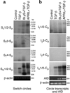
Figure 2. BLyS or APRIL and TGF-β induce CSR from Cμ to Cα
(a) IgD+ B cells were incubated with medium (control), TGF-β1, BLyS + TGF-β1 or APRIL + TGF-β1. Expression of Sα1/2Sμ and genomic β-actin after 4 days was assessed. (b) Expression of Iα1/2-Cμ, AID and β-actin transcripts; cells were treated as in a. One of three separate experiments that gave similar results is shown.
Figure 3
Figure 3. CSR is induced by BLyS, APRIL or CD40L but not by TNF-α or LT-α
(a) IgD+ B cells were incubated with medium alone (control), APRIL + IL-4, BLyS + IL-4, CD40L + IL-4, TNF-α + IL-4 or LT-α + IL-4. Expression of total Sγ-Sμ SCs, Sα1/2-Sμ SCs and β-actin after 4 days was assessed, (b) IgD+ B cells were incubated with IL-4 and 100, 10, 1 and 0.1 ng/ml of BLyS or APRIL. Expression of total Sγ-Sμ, after 4 days was assessed. One of three separate experiments that gave similar results is shown.
Figure 4
Figure 4. BLyS or APRIL up-regulate multiple germline IH-CH and mature VHDH-CH transcripts
(a) Germline IH-CH, mature VHDH-CH and β-actin transcripts from IgD+ B cells incubated for 4 days with IL-10, IL-4, BLyS or APRIL alone, BLyS + IL-10, BLyS + IL-4, APRIL+ IL-10 or APRIL + IL-4 were analyzed. The first lane of each panel shows a 100-bp DNA ladder, (b–d) NF-κB binding to DNA after incubation of IgD+ B cells with medium alone (control), BLyS or APRIL in the presence or absence of anti-BCR (b) and with or without a mouse control IgG1, TACI-Ig, BCMA-Ig or an antibody to soluble BLyS (sBLyS) (d). Arrows indicate p50–p65 and p50–c-Rel complexes, as identified by inhibition assays in which nuclear proteins from BLyS- or APRIL-activated B cells were first exposed to antibodies to p65, c-Rel, RelB and p50 or p52 and then incubated with the radiolabeled NF-κB–binding oligonucleotide (c). An irrelevant goat IgG was used as control. One of three separate experiments that gave similar results is shown.
Figure 5
Figure 5. BLyS or APRIL up-regulate both membrane-bound and secreted IgG and IgA
(a) IgD+ B cells were incubated with medium alone or BLyS and IL-4. Expression of surface IgM (sIgM), sIgD, sIgG and sIgA after 10 days was assessed. An isotype control is shown. Numbers indicate the percentages of class-switched B cells, (b) IgG and IgA secreted by IgD+ B cells incubated for 10 days with BLyS or APRIL + anti-BCR or with BLyS or APRIL + anti-BCR and IL-15 and in the presence or absence of TACI-Ig or BCMA-Ig was assessed. Mean ± s.d. data of one of three separate experiments that gave similar results are shown.
Figure 6
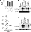
Figure 6. Activated DCs induce CSR through BLyS and APRIL
(a) DCs were incubated with medium alone, CD40L, IFN-α or IFN-γ. Expression of BLyS, APRIL and β-actin transcripts, and total BLyS, APRIL and actin proteins were assessed after 3 days. (b) Mean ± s.d. expression of sBLyS. (c) Expression of membrane-bound BLyS. Dotted and thick profiles depict control and BLyS antibodies, respectively, (d) IgD+ B cells were cocultured with unstimulated or stimulated DCs or epithelial cells (EPCs) in the presence or absence of a control mouse antibody, TACI-Ig, BCMA-Ig, CD40-Ig or an antibody to sBLyS. Iγ1/2-Cμ, Iγ3-Cμ and Iα1/2-Cμ CTs as well as AID and Igβ (CD79b, B cell–specific loading control) transcripts were amplified after 4 days.
Figure 7
Figure 7. Activated monocytes induce CSR through BLyS and APRIL
(a) Monocytes (Mos) were incubated with medium alone, IFN-γ, IFN-α or LPS for 3 days. Soluble and membrane-bound BLyS were measured after 3 days, (b) IgD+ B cells were incubated with resting or IFN-γ–activated monocytes in the presence or absence of a neutralizing antibodies to sBLyS, BCMA-Ig, TACI-Ig or CD40-Ig. Expression of Iα1/2-Cμ CTs and β-actin was assessed after 4 days, (c) IgD+ B cells were incubated with supernatants from monocytes (MoSup) cultured for 3 days alone or with IFN-γ in the presence or absence of a control mouse antibody, BCMA-Ig, TACI-Ig, CD40-Ig or an antibody to sBLyS. Iα1/2-Cμ CTs and β-actin were amplified after 4 days. One of three separate experiments that gave similar results is shown.
Figure 8
Figure 8. Activated DCs induce plasmacytoid differentiation through BLyS and APRIL
(a) BCR-activated IgD+ B cells were incubated with resting or CD40L-, IFN-α–or IFN-γ–activated DCs or epithelial cells in the presence or absence of a control antibody, TACI-Ig, BCMA-Ig, CD40-Ig or a neutralizing antibody to sBLyS or IL-15. Secreted IgG and IgA were measured after 10 days, (b–d), BCR-activated IgD+ B cells were incubated with IFN-α–activated epithelial cells, IL-15 and CD40-Ig (b), IFN-α–activated DCs, IL-15 and CD40-Ig (c), or IFN-α–activated DCs, IL-15 and BCMA-Ig (d). After 10 days, cells were stained with FITC-conjugated antibodies to IgA (green). Nuclear counterstaining was done with DAPI (blue, magnification:×1,000) and numbers indicate the percentages of cells expressing cytoplasmic IgA (e–g) IgD+ B cells in e–g were cultured for 10 days as in b–d, respectively. Numbers indicate percentages of CD38hiCD20lo and CD38hiCD 138+ plasmacytoid cells. Data are mean ± s.d. of one of three separate experiments that gave similar results.

Comment in

References

    1. Bassing CH, Swat W, Alt FW. The mechanism and regulation of chromosomal V(D)J recombination. Cell. 2002;109(Suppl):45–55. - PubMed
    1. Stavnezer J. Antibody class switching. Adv. Immunol. 1996;61:79–146. - PubMed
    1. Papavasiliou FN, Schatz DG. Somatic hypermutation of immunoglobulin genes: merging mechanisms for genetic diversity. Cell. 2002;109(Suppl):35–44. - PubMed
    1. Manis JP, Tian M, Alt FW. Mechanism and control of class-switch recombination. Trends Immunol. 2002;23:31–39. - PubMed
    1. MacLennan IC. Germinal centers. Annu. Rev. Immunol. 1994;12:117–139. - PubMed

Publication types

MeSH terms