

Astrocyte-endothelial interactions and blood-brain barrier permeability
- PMID: 12162730
- PMCID: PMC1570746
- DOI: 10.1046/j.1469-7580.2002.00064.x
Astrocyte-endothelial interactions and blood-brain barrier permeability
Abstract
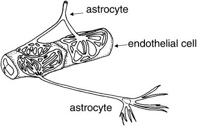
The blood-brain barrier (BBB) is formed by brain endothelial cells lining the cerebral microvasculature, and is an important mechanism for protecting the brain from fluctuations in plasma composition, and from circulating agents such as neurotransmitters and xenobiotics capable of disturbing neural function. The barrier also plays an important role in the homeostatic regulation of the brain microenvironment necessary for the stable and co-ordinated activity of neurones. The BBB phenotype develops under the influence of associated brain cells, especially astrocytic glia, and consists of more complex tight junctions than in other capillary endothelia, and a number of specific transport and enzyme systems which regulate molecular traffic across the endothelial cells. Transporters characteristic of the BBB phenotype include both uptake mechanisms (e.g. GLUT-1 glucose carrier, L1 amino acid transporter) and efflux transporters (e.g. P-glycoprotein). In addition to a role in long-term barrier induction and maintenance, astrocytes and other cells can release chemical factors that modulate endothelial permeability over a time-scale of seconds to minutes. Cell culture models, both primary and cell lines, have been used to investigate aspects of barrier induction and modulation. Conditioned medium taken from growing glial cells can reproduce some of the inductive effects, evidence for involvement of diffusible factors. However, for some features of endothelial differentiation and induction, the extracellular matrix plays an important role. Several candidate molecules have been identified, capable of mimicking aspects of glial-mediated barrier induction of brain endothelium; these include TGFbeta, GDNF, bFGF, IL-6 and steroids. In addition, factors secreted by brain endothelial cells including leukaemia inhibitory factor (LIF) have been shown to induce astrocytic differentiation. Thus endothelium and astrocytes are involved in two-way induction. Short-term modulation of brain endothelial permeability has been shown for a number of small chemical mediators produced by astrocytes and other nearby cell types. It is clear that endothelial cells are involved in both long- and short-term chemical communication with neighbouring cells, with the perivascular end feet of astrocytes being of particular importance. The role of barrier induction and modulation in normal physiology and in pathology is discussed.
Figures
References
-
- Abbott NJ, Revest PA. Control of brain endothelial permeability. Cerebrovasc. Brain Metab. Rev. 1991;3:39–72. - PubMed
-
- Abbott NJ, Romero IA. Transporting therapeutics across the blood–brain barrier. Mol. Med. Today. 1996;2:106–113. - PubMed
-
- Abbott NJ. Role of intracellular calcium in regulation of brain endothelial permeability. In: Pardridge WM, editor. Introduction to the Blood–Brain Barrier: Methodology and Biology. Cambridge, UK: Cambridge University Press; 1998. pp. 345–353.
-
- Allt G, Lawrenson JG. Is the pial microvessel a good model for blood–brain barrier studies? Brain Res. Rev. 1997;24:67–76. - PubMed
Publication types
MeSH terms
Grants and funding
LinkOut - more resources
Full Text Sources
Other Literature Sources
Miscellaneous
