. 2002 Aug;136(8):1093-7.
doi: 10.1038/sj.bjp.0704839.

Sphingosine 1-phosphate enhances spontaneous transmitter release at the frog neuromuscular junction

Affiliations

Sphingosine 1-phosphate enhances spontaneous transmitter release at the frog neuromuscular junction

Eugen Brailoiu et al. Br J Pharmacol. 2002 Aug.

Abstract

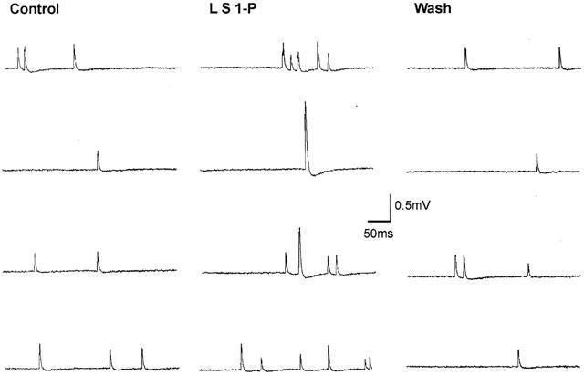
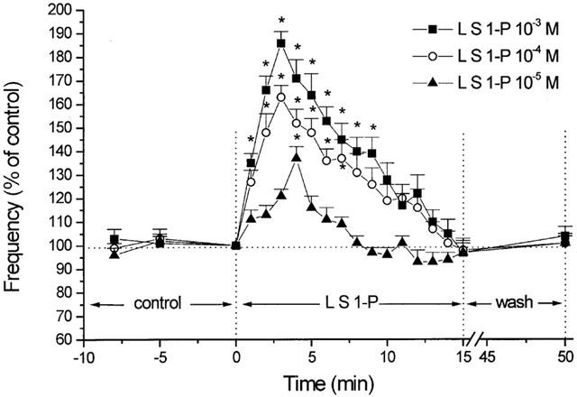
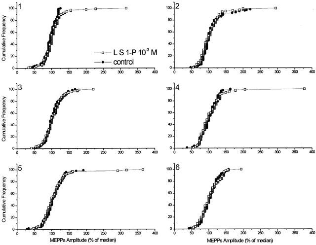
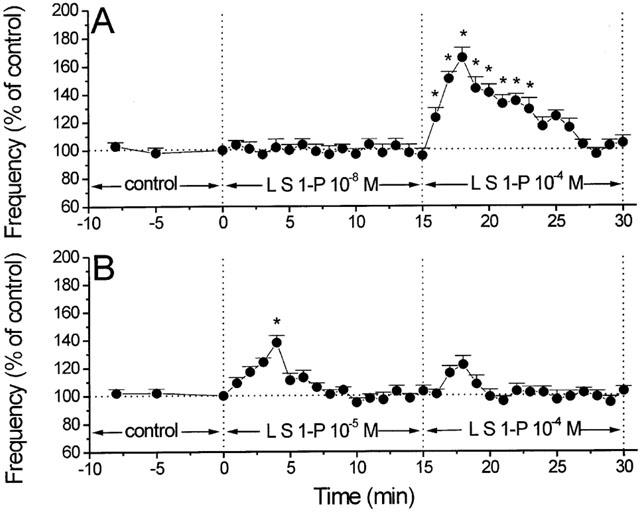
Intracellular recordings were made from isolated frog sciatic-sartorius nerve-muscle preparations, and the effects of sphingosine 1-phosphate (S1-P) on miniature endplate potentials (MEPPs) were studied. Extracellular application of S1-P (1 and 30 micro M) had no significant effects on the frequency and amplitude of MEPPs. Delivery into nerve terminals by liposomes containing 10(-5), 10(-4) or 10(-3) M S1-P was associated with a concentration-dependent increase in MEPP frequency of 37, 63 and 86%. The per cent of median MEPP amplitude was not significantly changed, but there was an increase in the number of 'giant' MEPPs. Pre-exposure of the preparations to S1-P 10(-5) but not 10(-8) M entrapped in liposomes for 15 min blocked the effects of subsequent superfusion of S1-P (10(-4) M)-filled liposomes on MEPP frequency. Thus, intracellular S1-P receptors seem to undergo 'desensitization' to higher concentrations of S1-P. The result provides the first evidence that S1-P acting intracellularly but not extracellularly enhances spontaneous transmitter release at the frog neuromuscular junction.


Figures

Figure 1
Figure 1
Sample recordings of miniature endplate potentials (MEPPs) in normal Ringer solution before, during, and after superfusion of S1-P (10−3 M) entrapped in liposomes. Note the increase in MEPP frequency and the presence of ‘giant' MEPPs during S1-P superfusion.
Figure 2
Figure 2
Percent changes in MEPP frequency as a function of time. The effects of 10−5, 10−4 and 10−3 M S1-P delivered by liposomes (L S1-P) on MEPP frequency are superimposed for comparison. The peak effect occurs at 3 min for 10−4 and 10−3 M, and at 4 min for 10−5 M S1-P. MEPP frequency 100%=0.974 s−1 (10−5 M S1-P), 1.12 s−1 (10−4 M S1-P) and 0.983 s−1 (10−3 M S1-P). Note that the MEPP frequency return to control level, e.g., at time=8 min for S1-P 10−5 M. Each point represents the mean from six different experiments. Asterisks denote statistically significant differences (P<0.05) from control.
Figure 3
Figure 3
Histogram analysis (cumulative frequency) of the changes in MEPP amplitude distribution before (control) and after S1-P delivered by liposomes (L S1-P). For each of the six single experiments, MEPP amplitudes at time=0 min (pre-exposure) and time=3 min (post-exposure) are expressed as a per cent of the median amplitude (100 samples each). Histograms reveal no change in the shape of the unimodal amplitude-frequency distribution of MEPPs (exp. 1 median=0.348 mV; exp. 2 median=0.317 mV; exp. 3 median=0.392 mV; exp. 4 median=0.329 mV; exp. 5 median=0.377 mV; exp. 6 median=0.366 mV) before and after administration of liposomes containing 10−3 M S1-P. Results indicate no significant changes in the median MEPP amplitude before and after S1-P treatment; however, there is an increase in the number of gMEPPs after S1-P treatment.
Figure 4
Figure 4
Responses of second liposomal delivery of S1-P following a low and higher concentration of liposomal delivery of S1-P on MEPP frequency. (A) Perfusion with liposomes containing S1-P 10−8 M (L S1-P 10−8 M) had no significant effect on MEPP frequency. A subsequent administration of S1-P 10−4 M-filled liposomes (L S1-P 10−4 M) increased the MEPP frequency to a degree similar to that of muscle preparations treated with S1-P 10−4 M-filled liposomes alone (P>0.05) (n=6). Control MEPP frequency (min 0)=1.17 s−1. (B) Administration of S1-P 10−4 M-filled liposomes (L S1-P 10−4 M) to preparations pre-exposed to S1-P 10−5 entrapped liposomes (L S1-P 10−5 M) induced no significant changes in MEPP frequency (n=6). Control MEPP frequency (min 0)=0.88 s−1. In all cases, asterisks denote statistically significant differences (P<0.05) from control.

References

    1. ALEMANY R., KLEUSER B., RUWISCH L., DANNEBERG K., LASS H., HASHEMI R., SPIEGEL S., JAKOBS K.H., MEYER ZU HERINGDORF D. Depolarisation induces rapid and transient formation of intracellular sphingosine-1-phosphate. FEBS Lett. 2001;509:239–244. - PubMed
    1. ALKADHI K.A. Emetine increases giant miniature endplate potential population at the frog neuromuscular junction. Brain Res. 1988;447:293–298. - PubMed
    1. BRAILOIU E., MIYAMOTO M.D. Inositol phosphates and cyclic adenosine diphosphate-ribose increase quantal transmitter release at frog nerve terminals: possible involvement of smooth endoplasmic reticulum. Neurosci. 2000;95:927–931. - PubMed
    1. BRAILOIU E., MIYAMOTO M.D., DUN N.J. Nicotinic acid adenine dinucleotide phosphate enhances quantal neurosecretion at frog neuromuscular junction: possible action on synaptic vesicles in the releasable pool. Mol. Pharmacol. 2001;60:718–724. - PubMed
    1. CALUPCA M.A., PRIOR C., MERRIAM L.A., HENDRICKS G.M., PARSONS R.L. Presynaptic function is altered in snake K+-depolarized motor nerve terminals containing compromised mitochondria. J. Physiol. (Lond.) 2001;532:217–227. - PMC - PubMed

Publication types

MeSH terms

LinkOut - more resources