

Studies of GABA(B) receptors labelled with [(3)H]-CGP62349 in hippocampus resected from patients with temporal lobe epilepsy
- PMID: 12163342
- PMCID: PMC1573440
- DOI: 10.1038/sj.bjp.0704812
Studies of GABA(B) receptors labelled with [(3)H]-CGP62349 in hippocampus resected from patients with temporal lobe epilepsy
Abstract
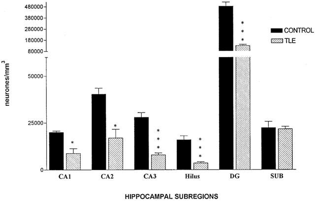
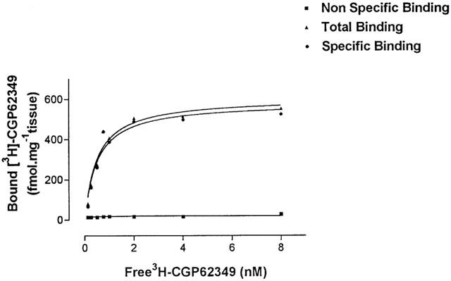
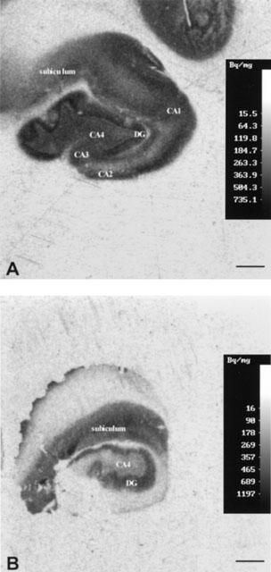
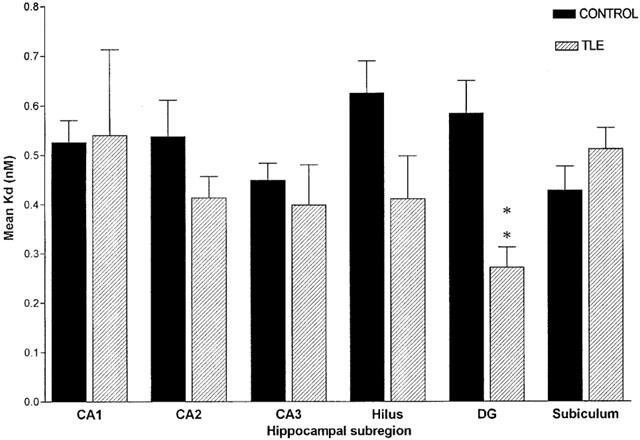
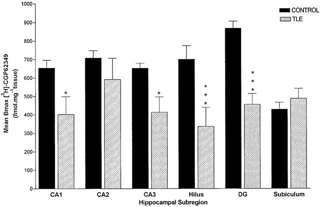
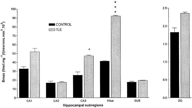
1 The aim of this study was to investigate the binding of a novel GABA(B) receptor radioligand, [(3)H]-CGP62349, to human post-mortem control and epileptic hippocampal sections using quantitative receptor autoradiography. Utilizing human control hippocampal sections it was shown that [(3)H]-CGP62349 bound with high affinity (K(D) 0.5 nM) to this tissue. 2 Hippocampal slices from surgical specimens obtained from patients with hippocampal sclerosis (HS) and temporal lobe epilepsy (TLE) were compared with neurologically normal post-mortem control subjects for neuropathology and GABA(B) receptor density and affinity. Neuronal loss was observed in most of the hippocampal subregions, but in the subiculum no significant difference was detected. 3 The localization of GABA(B) receptors with the antagonist [(3)H]-CGP62349 in human control hippocampal sections supported and extended earlier studies using the agonist ligand [(3)H]-GABA. 4 The kinetics of binding to the GABA(B) receptor in human hippocampus using this novel compound was comparable to previous data obtained in rat hippocampal membranes. 5 GABA(B) receptor density (B(max)) was significantly reduced in CA3, hilus, and dentate gyrus (DG); the affinity was increased exclusively in DG. The trend is identical in all the hippocampal subregions with the agonist and the antagonist, although significant differences with the antagonist where recorded in CA3 and hilus, whereas with the agonist a significant reduction was reported in all of the hippocampal subfields. 6 GABA(B) receptor expression per remaining neuron appeared significantly increased in CA3 and hilus. These results suggest altered GABA(B) receptor function may occur in human TLE, possibly as a result of synaptic reorganization, and may contribute to epileptogenesis.
Figures
References
-
- ABERCOMBIE M. Estimation of nuclear populations from microtome sections. Anat. Rec. 1946;94:239–247. - PubMed
-
- AMARAL D.G., INSAUSTI R.Hippocampal formation The human nervous system 1990New York: Accademic Press; 711–752.ed. Paxinios, G pp
-
- ANDRADE R., MALENKA R.C., NICOLL R.A. A G protein couples serotonin and GABAB receptors to the same channel in hippocampus. Science. 1986;234:1261–1265. - PubMed
-
- ASPRODINI E.K., RAINNIE D.G., SHINNICK-GALLAGHER P. Epileptogenesis reduces the sensitivity of presynaptic GABAB receptors on glutamatergic afferents in the amigdala. J. Pharmacol. Exp. Ther. 1992;262:1011–1021. - PubMed
-
- BABB T.L., BROWN W.J., PRETORIUS J.K., DAVENPORT C., LIEB J.P., CRANDALL P.H. Temporal lobe volumetric cell densities in temporal lobe epilepsy. Epilepsia. 1984;25:729–793. - PubMed
Publication types
MeSH terms
Substances
LinkOut - more resources
Full Text Sources
Miscellaneous
